Copyright
©The Author(s) 2024.
World J Clin Cases. Jun 16, 2024; 12(17): 3243-3252
Published online Jun 16, 2024. doi: 10.12998/wjcc.v12.i17.3243
Published online Jun 16, 2024. doi: 10.12998/wjcc.v12.i17.3243
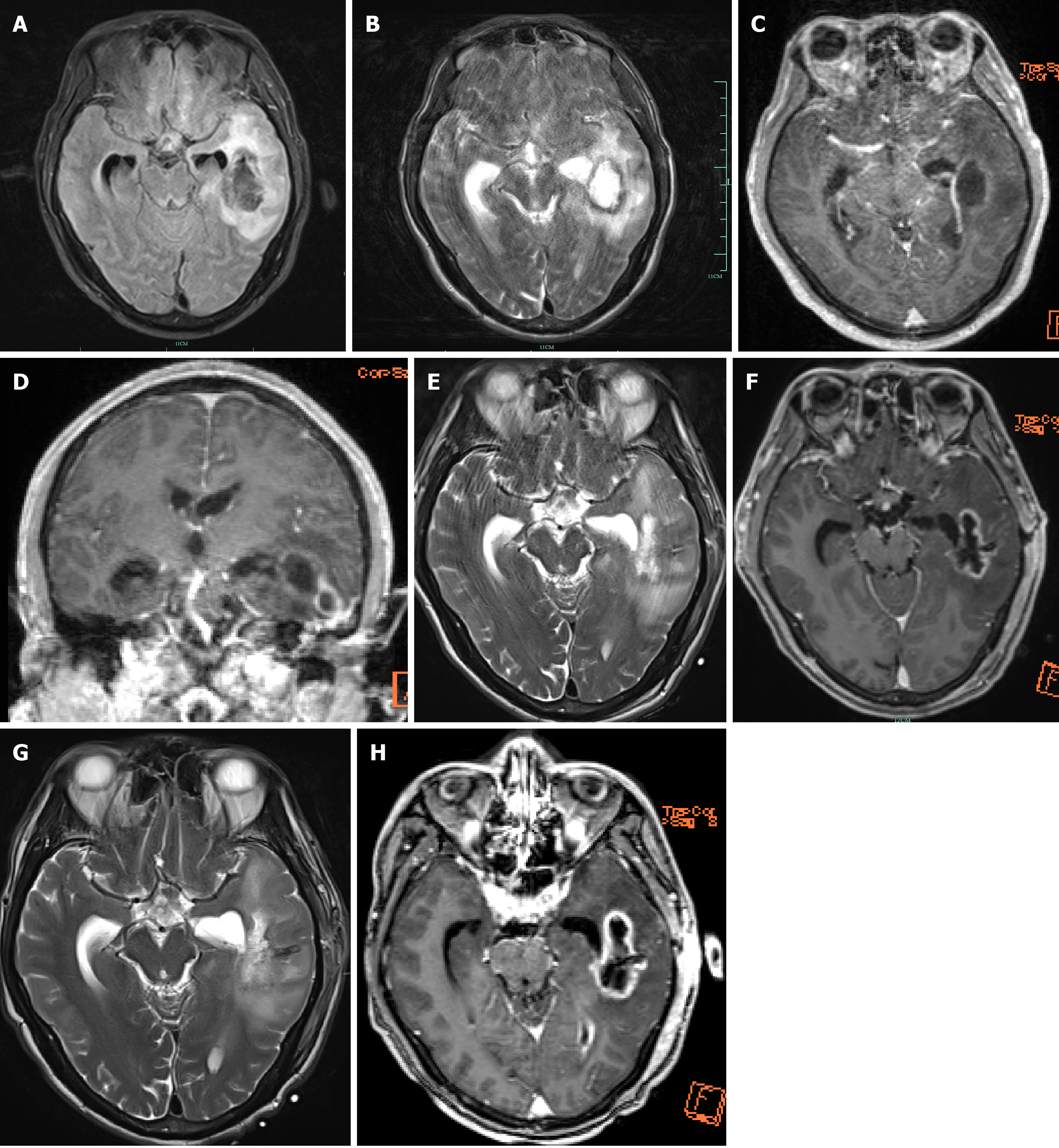
Figure 3 The lesion in the left temporal lobe with marked surrounding edema.
After enhancement, the lesion shows ring-like enhancement, the left lateral ventricle is compressed and narrowed, the midline structures are slightly shifted to the right, and purulent meningitis has ruptured into the ventricular system. Re-examination 6 days post-surgery, shows that compared to the previous image from September 06 2021, the left temporal lobe abscess has decreased in size, and purulent meningitis has ruptured into the ventricular system. Twenty days post-surgery, re-examination shows that the abscess cavity has significantly reduced in size, the surrounding edema has reduced, and the purulent meningitis has ameliorated compared to the initial status. The characteristic ring enhancement of a brain abscess is a primary feature, and the differ from brain tumors in that brain tumors rarely show this characteristic ring enhancement. Additionally, the internal enhancement of brain tumors is often unevenly distributed with signals, and surrounding tumor blood vessels are visible. T2 imaging reveals that they often appear as curve-shaped or pinpoint-shaped low-signal shadows due to the flow void effect. A-D: Magnetic resonance imaging (MRI) T2 suppressed water imaging, MRI T2 scans, both, MRI enhanced scans (C and D), respectively, from September 6 2021; E and F: MRI T2 scans, MRI enhanced scans, respectively, from September 12 2021; H and G: MRI T2 scans, MRI enhanced scans, respectively, from September 16 2021.
- Citation: Tan SD, Li MH. Brain abscess caused by Streptococcus anginosus group: Three case reports. World J Clin Cases 2024; 12(17): 3243-3252
- URL: https://www.wjgnet.com/2307-8960/full/v12/i17/3243.htm
- DOI: https://dx.doi.org/10.12998/wjcc.v12.i17.3243


