Copyright
©The Author(s) 2024.
World J Clin Cases. Jun 16, 2024; 12(17): 3243-3252
Published online Jun 16, 2024. doi: 10.12998/wjcc.v12.i17.3243
Published online Jun 16, 2024. doi: 10.12998/wjcc.v12.i17.3243
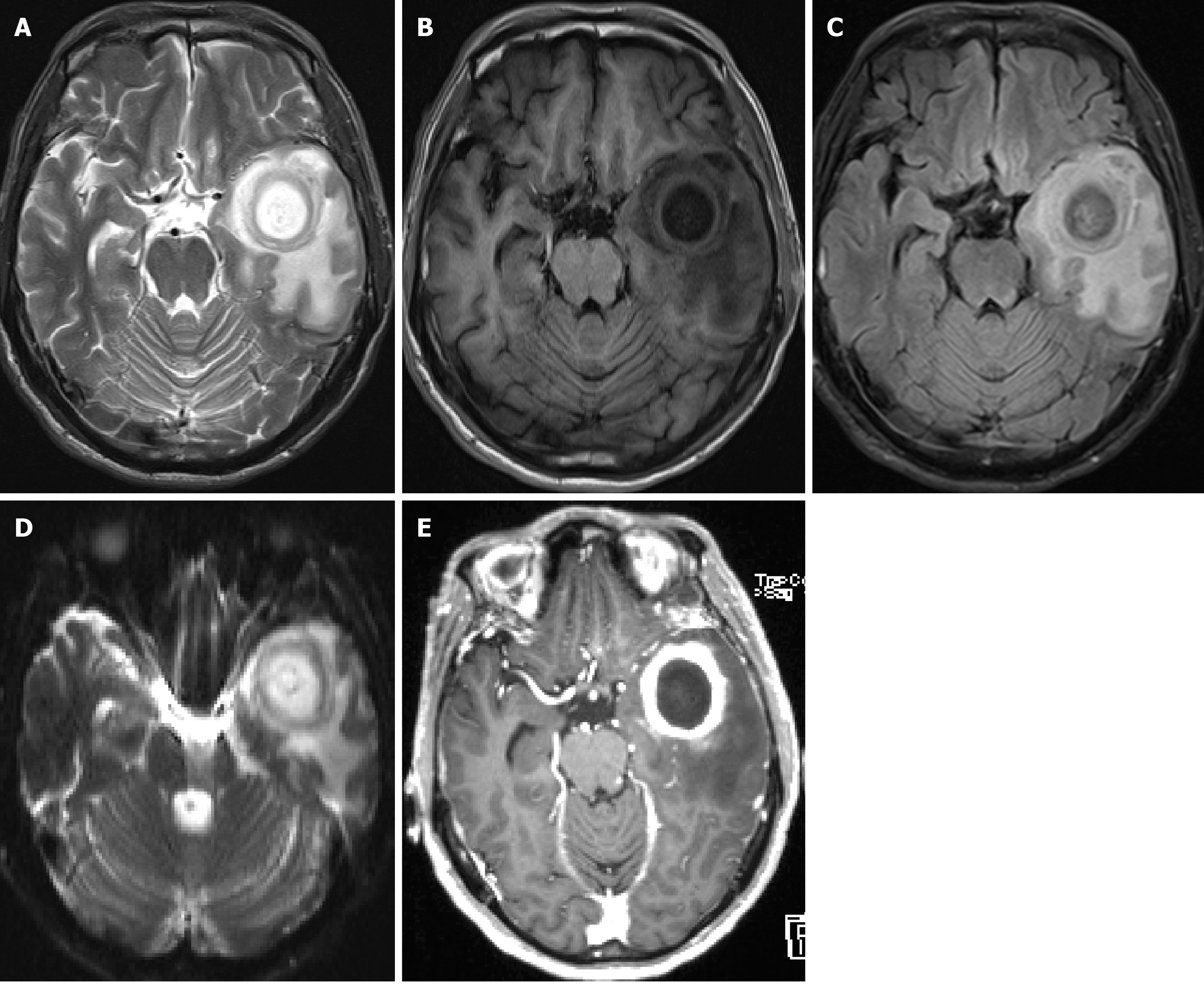
Figure 2 The preoperative magnetic resonance imaging scans from March 30 2017.
An oval-shaped mass in the left temporal lobe with visible patchy edema surrounding it. The contrast-enhanced scan shows prominent ring enhancement, with fluffy and small patchy enhancements around the ring. The left lateral ventricle and midline structures are slightly compressed and shifted. A brain abscess in the left temporal lobe is highly probable, however, tumor-like lesions cannot be completely ruled out due to the presence of fluffy enhancements. Diffusion-weighted imaging (DWI) shows that the edema around brain abscess lesions can vary from mild to severe, showing high or relatively high signals, while the edema around necrotic brain tumors is generally severe and shows high signals. Finally, the contrast-enhanced scan shows that the wall of a brain abscess is regular and relatively uniform, while the wall of a necrotic brain tumor is uneven in thickness, with mural nodules, showing a flower ring-like or annular enhancement. A: Magnetic resonance imaging (MRI) T2; B: MRI T1; C: MRI T2 water suppression imaging; D: MRI DWI scans; E: MRI contrast-enhanced scans.
- Citation: Tan SD, Li MH. Brain abscess caused by Streptococcus anginosus group: Three case reports. World J Clin Cases 2024; 12(17): 3243-3252
- URL: https://www.wjgnet.com/2307-8960/full/v12/i17/3243.htm
- DOI: https://dx.doi.org/10.12998/wjcc.v12.i17.3243


