Copyright
©The Author(s) 2024.
World J Clin Cases. Jun 16, 2024; 12(17): 3243-3252
Published online Jun 16, 2024. doi: 10.12998/wjcc.v12.i17.3243
Published online Jun 16, 2024. doi: 10.12998/wjcc.v12.i17.3243
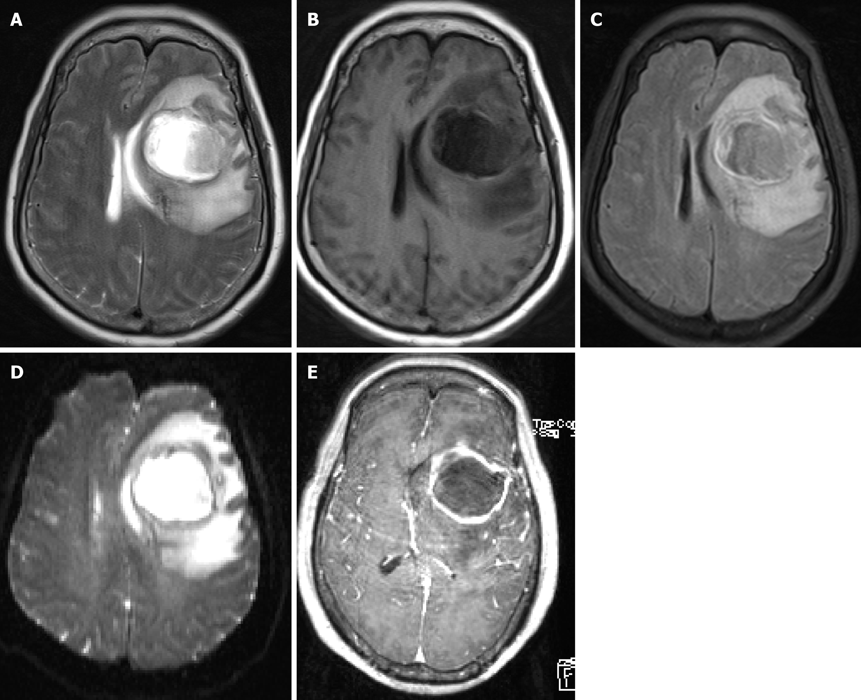
Figure 1 The preoperative magnetic resonance imaging scan from April 16 2019.
A space-occupying lesion in the left frontal lobe with extensive surrounding edema, showing marked ring enhancement on the enhanced scan. The left lateral ventricle is compressed, and the midline is shifted to the right. A brain abscess with ring enhancement is the primary diagnosis. A: Magnetic resonance imaging (MRI) T2; B: MRI T1; C: MRI T2 suppressed water imaging; D: MRI diffusion-weighted imaging scans; E: MRI enhanced scans.
- Citation: Tan SD, Li MH. Brain abscess caused by Streptococcus anginosus group: Three case reports. World J Clin Cases 2024; 12(17): 3243-3252
- URL: https://www.wjgnet.com/2307-8960/full/v12/i17/3243.htm
- DOI: https://dx.doi.org/10.12998/wjcc.v12.i17.3243


