Copyright
©The Author(s) 2024.
World J Clin Cases. Jun 16, 2024; 12(17): 3226-3234
Published online Jun 16, 2024. doi: 10.12998/wjcc.v12.i17.3226
Published online Jun 16, 2024. doi: 10.12998/wjcc.v12.i17.3226
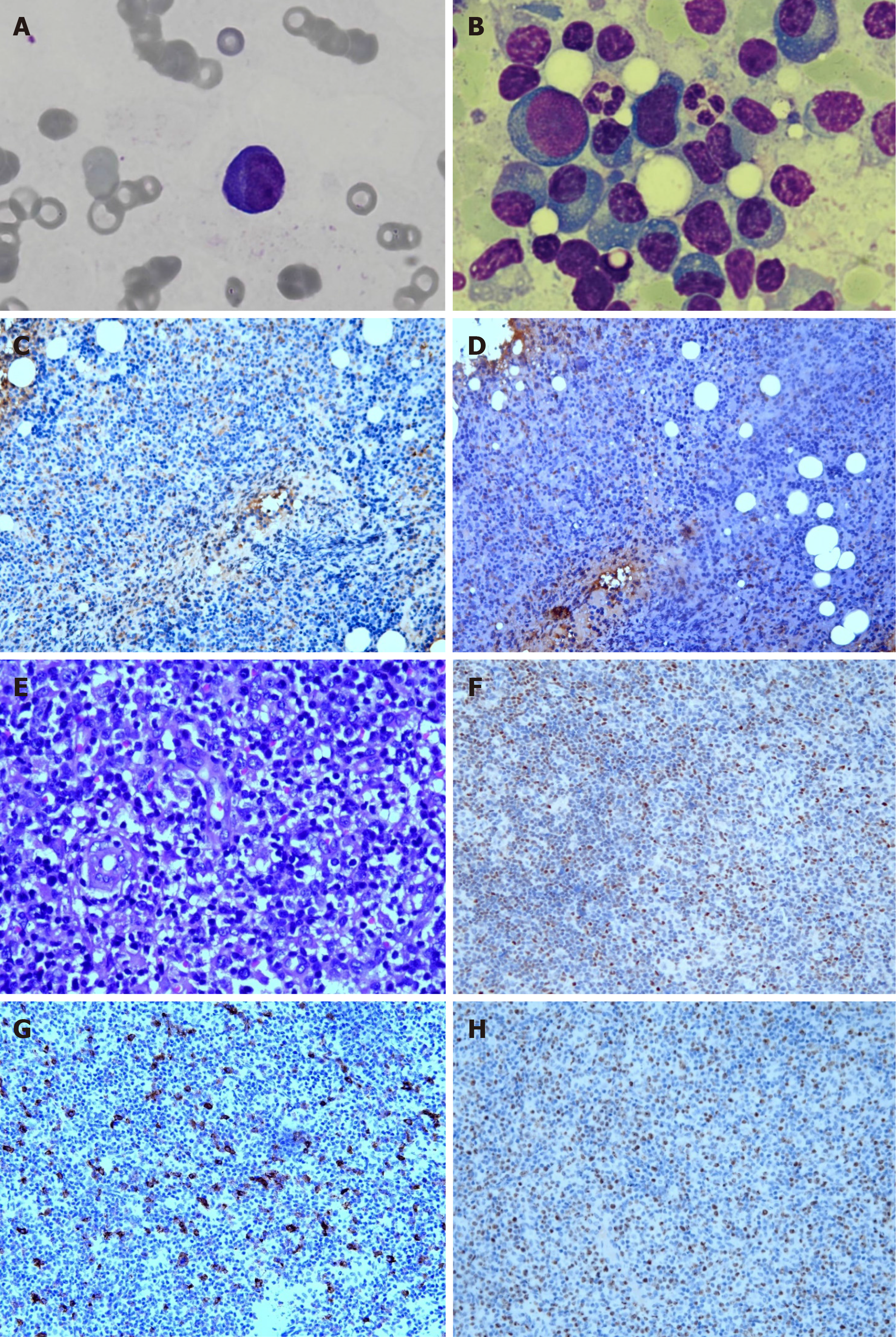
Figure 2 Microscopic evaluation of the lesion.
A: A plasma cell and rouleaux formation in peripheral blood smear (magnification, 1000 ×); B: Plasmacytosis with atypical and immature plasma cells in bone marrow smear (magnification, 1000 ×); C: Immunohistochemistry (IHC) results for kappa show positivity in bone marrow (magnification, 200 ×); D: IHC results for lambda show positivity in bone marrow (magnification, 200 ×); E: Small- to medium-sized atypical lymphocytes and follicular dendritic cells with clear cytoplasm and capillaries with enlarged endothelial cells are observed using hematoxylin and eosin staining (magnification, 400 ×); F: IHC results for BCL-6 show positivity (magnification, 100 ×); G: IHC results for CD30 show positivity (magnification, 100 ×); H: IHC results for Ki-67 show positivity (magnification, 100 ×).
- Citation: Lin CC, Lee HL, Chuo HY, Chen TA, Liu MY, Chen LM. Plasmacytosis mimicking multiple myeloma in angioimmunoblastic T-cell lymphoma: A case report and review of literature. World J Clin Cases 2024; 12(17): 3226-3234
- URL: https://www.wjgnet.com/2307-8960/full/v12/i17/3226.htm
- DOI: https://dx.doi.org/10.12998/wjcc.v12.i17.3226


