Copyright
©The Author(s) 2024.
World J Clin Cases. Jun 16, 2024; 12(17): 3206-3213
Published online Jun 16, 2024. doi: 10.12998/wjcc.v12.i17.3206
Published online Jun 16, 2024. doi: 10.12998/wjcc.v12.i17.3206
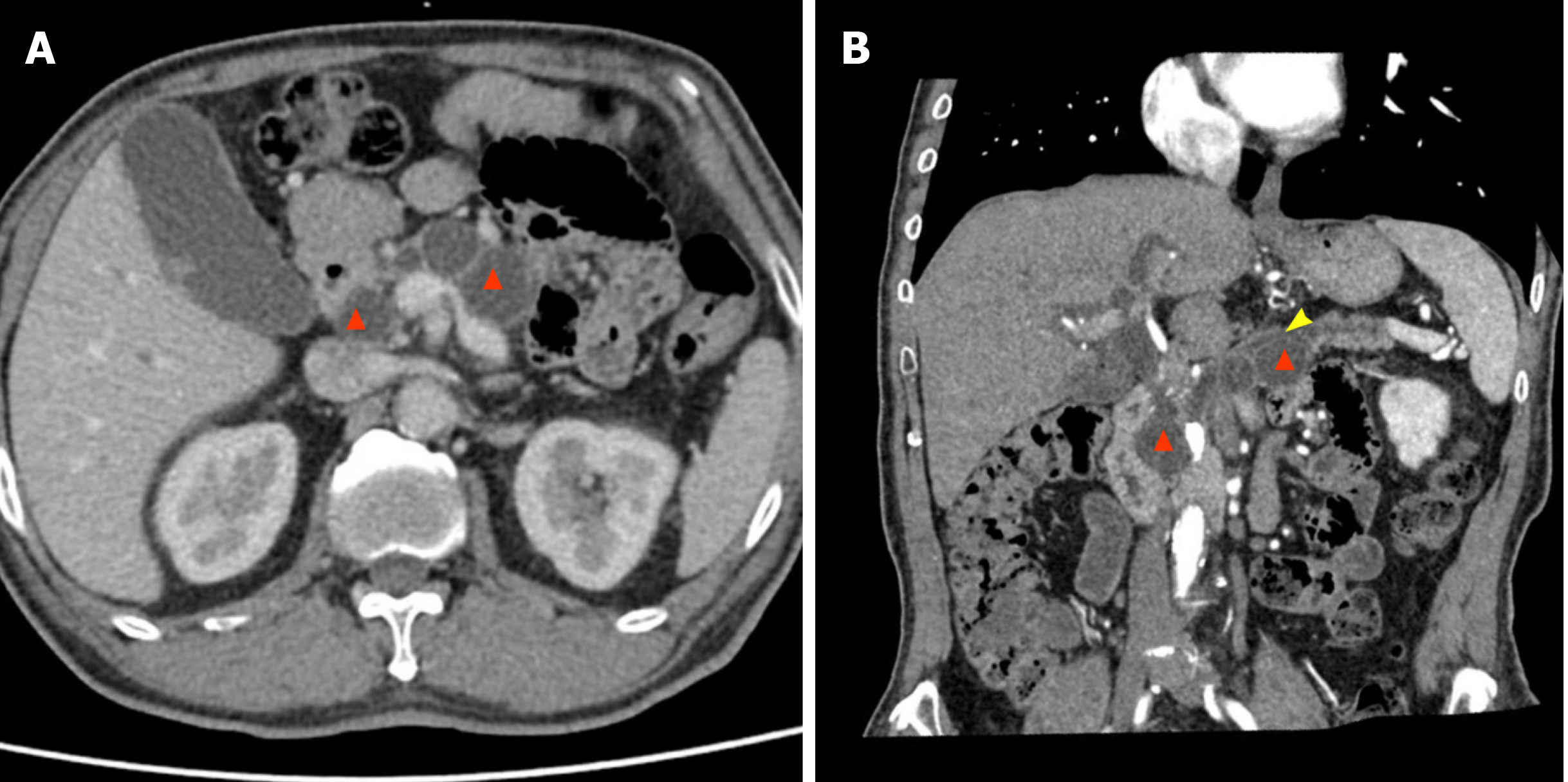
Figure 2 Enhanced abdominal computed tomography scan of case 2.
Multiple cystic low-density shadows are visible in the pancreatic head and body, with pancreatic parenchymal atrophy and dilation of the main pancreatic duct. Red triangle: Multiple cystic masses in the pancreatic head and body; Yellow arrow: Dilation of the main pancreatic duct. A: Axial phase; B: Reconstruction of the biliary and pancreatic duct system.
- Citation: Sun MQ, Kang XM, He XD, Han XL. Laparoscopic spleen-preserving total pancreatectomy for the treatment of low-grade malignant pancreatic tumors: Two case reports and review of literature. World J Clin Cases 2024; 12(17): 3206-3213
- URL: https://www.wjgnet.com/2307-8960/full/v12/i17/3206.htm
- DOI: https://dx.doi.org/10.12998/wjcc.v12.i17.3206


