Copyright
©The Author(s) 2024.
World J Clin Cases. Jun 16, 2024; 12(17): 3200-3205
Published online Jun 16, 2024. doi: 10.12998/wjcc.v12.i17.3200
Published online Jun 16, 2024. doi: 10.12998/wjcc.v12.i17.3200
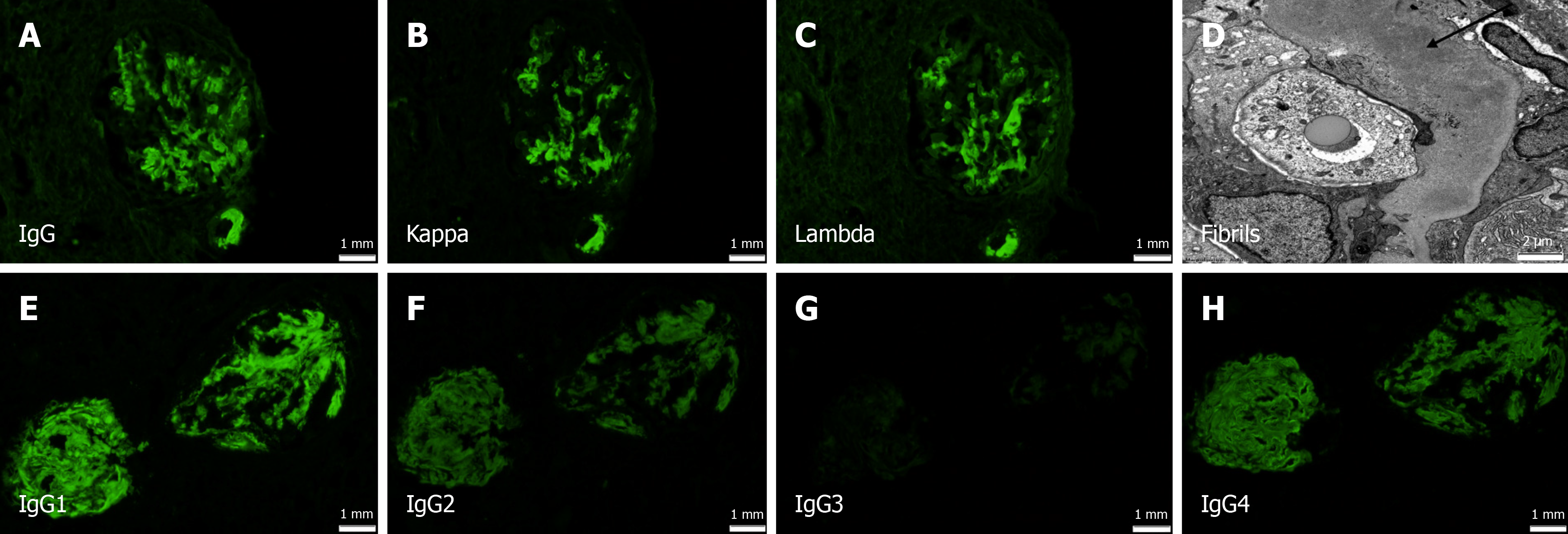
Figure 1 Direct immunofluorescence on frozen tissue along with electron microscopy.
A-C: Immunoglobulin gamma (IgG), kappa and lambda, smudgy predominantly mesangial staining; D: Ultrastructural examination shows randomly arranged fibrils in the glomeruli, measuring 10 nm in mean diameter (uranyl acetate lead citrate fixation); E-H: Direct immunofluorescence (DIF) for IgG subclasses demonstrated predominantly IgG1 but also mild to moderate IgG2 and IgG4, appears polytypic. IgG3 is negative (DIF images 40 ×).
- Citation: Chow MBCY, Bushrow L, Siddiqui I, Chiu A, Hamirani M, Satoskar AA. Congophilic fibrils in the glomeruli with polyclonal immunoglobulin gamma staining - another cause for diagnostic overlap: A case report. World J Clin Cases 2024; 12(17): 3200-3205
- URL: https://www.wjgnet.com/2307-8960/full/v12/i17/3200.htm
- DOI: https://dx.doi.org/10.12998/wjcc.v12.i17.3200


