Copyright
©The Author(s) 2024.
World J Clin Cases. Jun 16, 2024; 12(17): 3177-3182
Published online Jun 16, 2024. doi: 10.12998/wjcc.v12.i17.3177
Published online Jun 16, 2024. doi: 10.12998/wjcc.v12.i17.3177
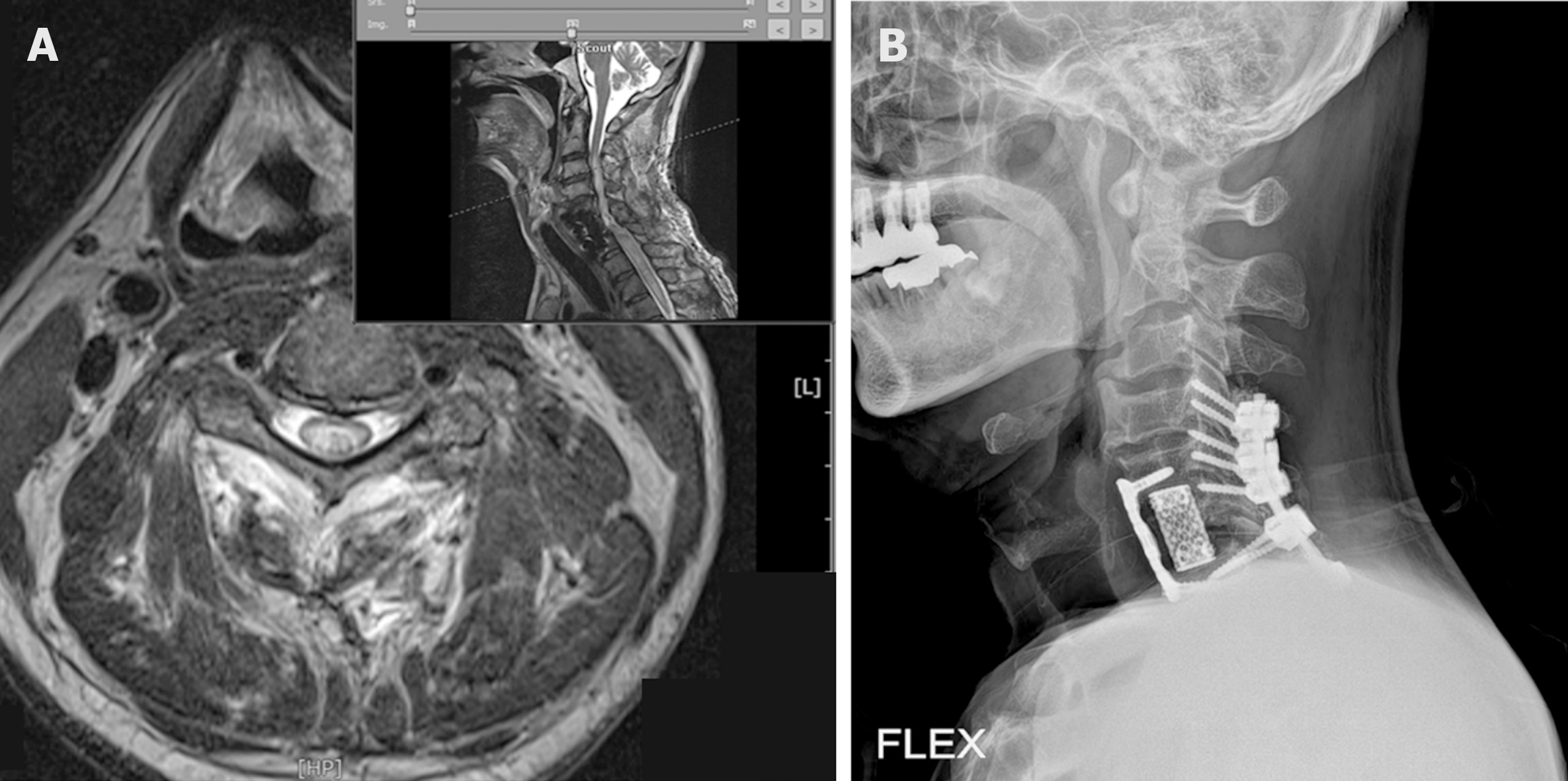
Figure 2 Cervical magnetic resonance imaging study.
A and B: Showing diffuse intramedullary increased T2 signal lesion from C2-3 to upper thoracic level (A), and cervical spine lateral view (B).
- Citation: Park HS, Kim JH. Effect of transcranial direct current stimulation on supernumerary phantom limb pain in spinal cord injured patient: A case report. World J Clin Cases 2024; 12(17): 3177-3182
- URL: https://www.wjgnet.com/2307-8960/full/v12/i17/3177.htm
- DOI: https://dx.doi.org/10.12998/wjcc.v12.i17.3177


