Copyright
©The Author(s) 2024.
World J Clin Cases. Jun 16, 2024; 12(17): 3156-3160
Published online Jun 16, 2024. doi: 10.12998/wjcc.v12.i17.3156
Published online Jun 16, 2024. doi: 10.12998/wjcc.v12.i17.3156
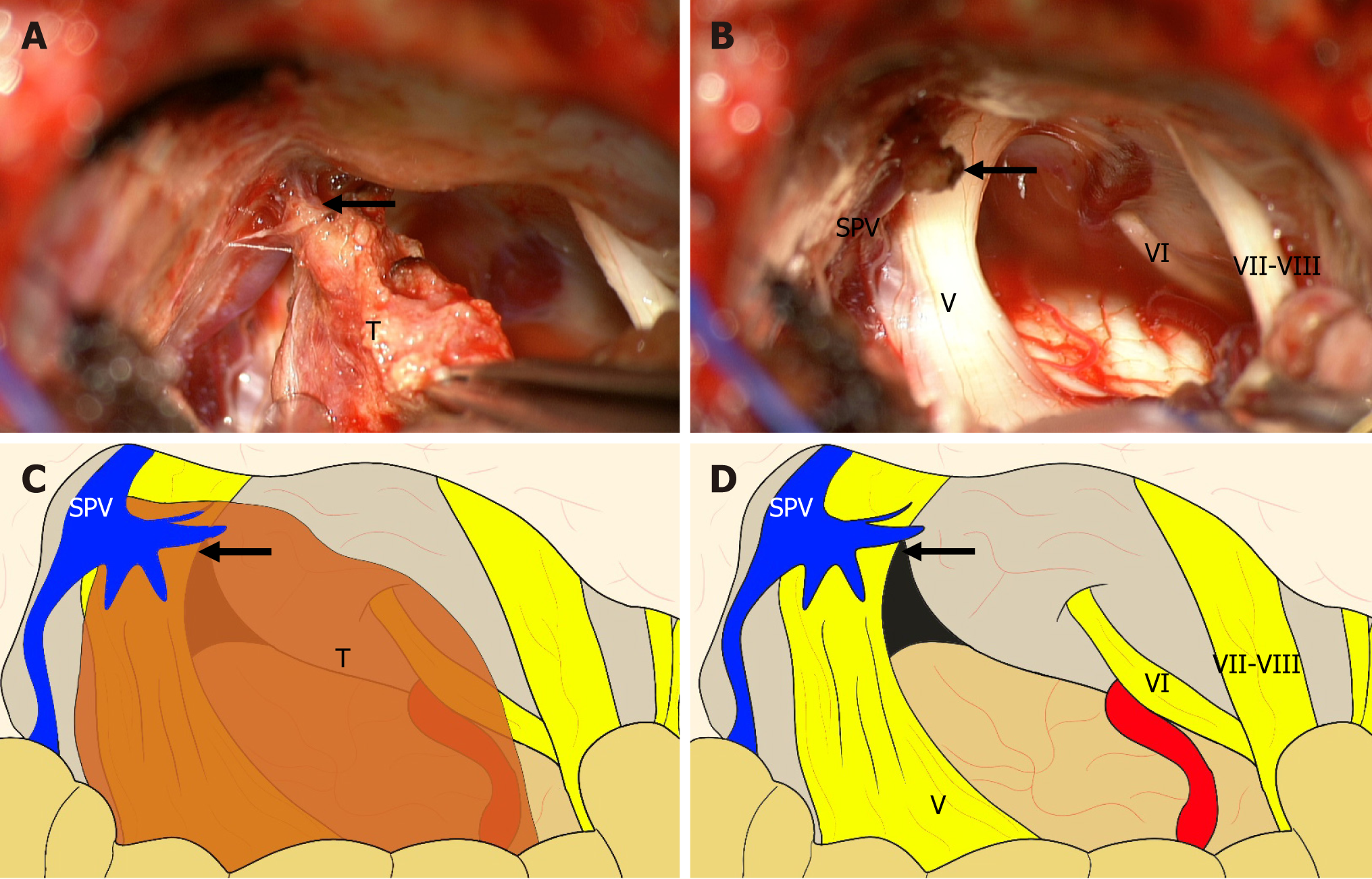
Figure 2 Intraoperative microscopic and schematic images.
A: The tumor was firmly adherent to the superior petrosal vein without dural attachment to the tentorium or petrous pyramid; B: The origin site was cauterized and resected to completely remove the tumor; C: The simplified schematic image of the intraoperative microscopic image A; D: The simplified schematic image of the intraoperative microscopic image B. Arrow: Origin site; T: Tumor; SPV: Superior petrosal vein; V: Trigeminal nerve; VI: Abducens nerve; VII-VIII: Facial-vestibulocochlear nerve complex.
- Citation: Kim YJ, Jung S, Jung TY, Moon KS, Kim IY. Meningioma originating from the superior petrosal vein without dural attachment: A case report. World J Clin Cases 2024; 12(17): 3156-3160
- URL: https://www.wjgnet.com/2307-8960/full/v12/i17/3156.htm
- DOI: https://dx.doi.org/10.12998/wjcc.v12.i17.3156


