Copyright
©The Author(s) 2024.
World J Clin Cases. Jun 16, 2024; 12(17): 3156-3160
Published online Jun 16, 2024. doi: 10.12998/wjcc.v12.i17.3156
Published online Jun 16, 2024. doi: 10.12998/wjcc.v12.i17.3156
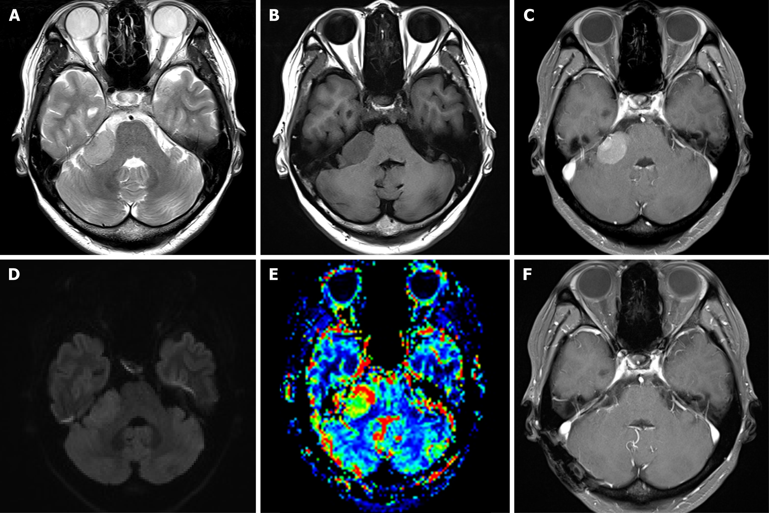
Figure 1 Preoperative imaging.
A and B: An extra-axial mass in the right cerebellopontine angle with isointensity on T2-wieghted image and iso-/hypointense signal mass on the T1-weighted image; C: Magnetic resonance imaging with gadolinium enhancement revealing a homogeneously enhancing mass without dural tail sign; D: Diffusion-weighted imaging; E: The corrected relative cerebral blood volume map showed high blood volume in the areas of the mass; F: Postoperative gadolinium-enhanced axial T1-weighted image shows no evidence of residual tumor.
- Citation: Kim YJ, Jung S, Jung TY, Moon KS, Kim IY. Meningioma originating from the superior petrosal vein without dural attachment: A case report. World J Clin Cases 2024; 12(17): 3156-3160
- URL: https://www.wjgnet.com/2307-8960/full/v12/i17/3156.htm
- DOI: https://dx.doi.org/10.12998/wjcc.v12.i17.3156


