Copyright
©The Author(s) 2024.
World J Clin Cases. Jun 6, 2024; 12(16): 2894-2903
Published online Jun 6, 2024. doi: 10.12998/wjcc.v12.i16.2894
Published online Jun 6, 2024. doi: 10.12998/wjcc.v12.i16.2894
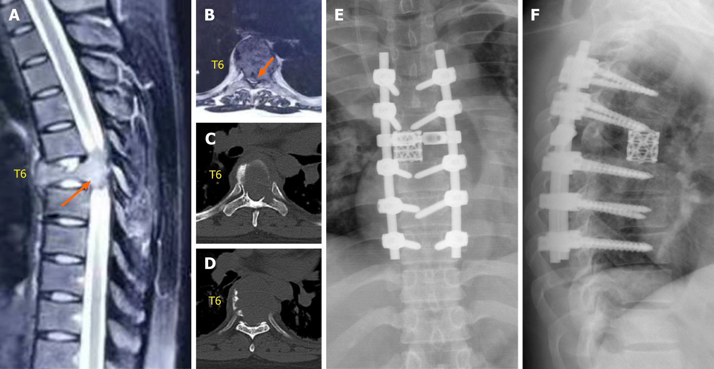
Figure 2 Preoperative and postoperative imaging of the first emergency total enbloc spondylectomy.
A and B: Magnetic resonance imaging before the first emergency total en bloc spondylectomy (arrow, tumor); C and D: Computed tomography before the first emergency total en bloc spondylectomy; E and F: Anteroposterior and lateral radiographs showing a good implant position at 1 wk after the first total en bloc spondylectomy.
- Citation: Liang HF, Xu H, Zhan MN, Xiao J, Li J, Fei QM. Thoracic giant cell tumor after two total en bloc spondylectomies including one emergency surgery: A case report. World J Clin Cases 2024; 12(16): 2894-2903
- URL: https://www.wjgnet.com/2307-8960/full/v12/i16/2894.htm
- DOI: https://dx.doi.org/10.12998/wjcc.v12.i16.2894


