Copyright
©The Author(s) 2024.
World J Clin Cases. Jun 6, 2024; 12(16): 2887-2893
Published online Jun 6, 2024. doi: 10.12998/wjcc.v12.i16.2887
Published online Jun 6, 2024. doi: 10.12998/wjcc.v12.i16.2887
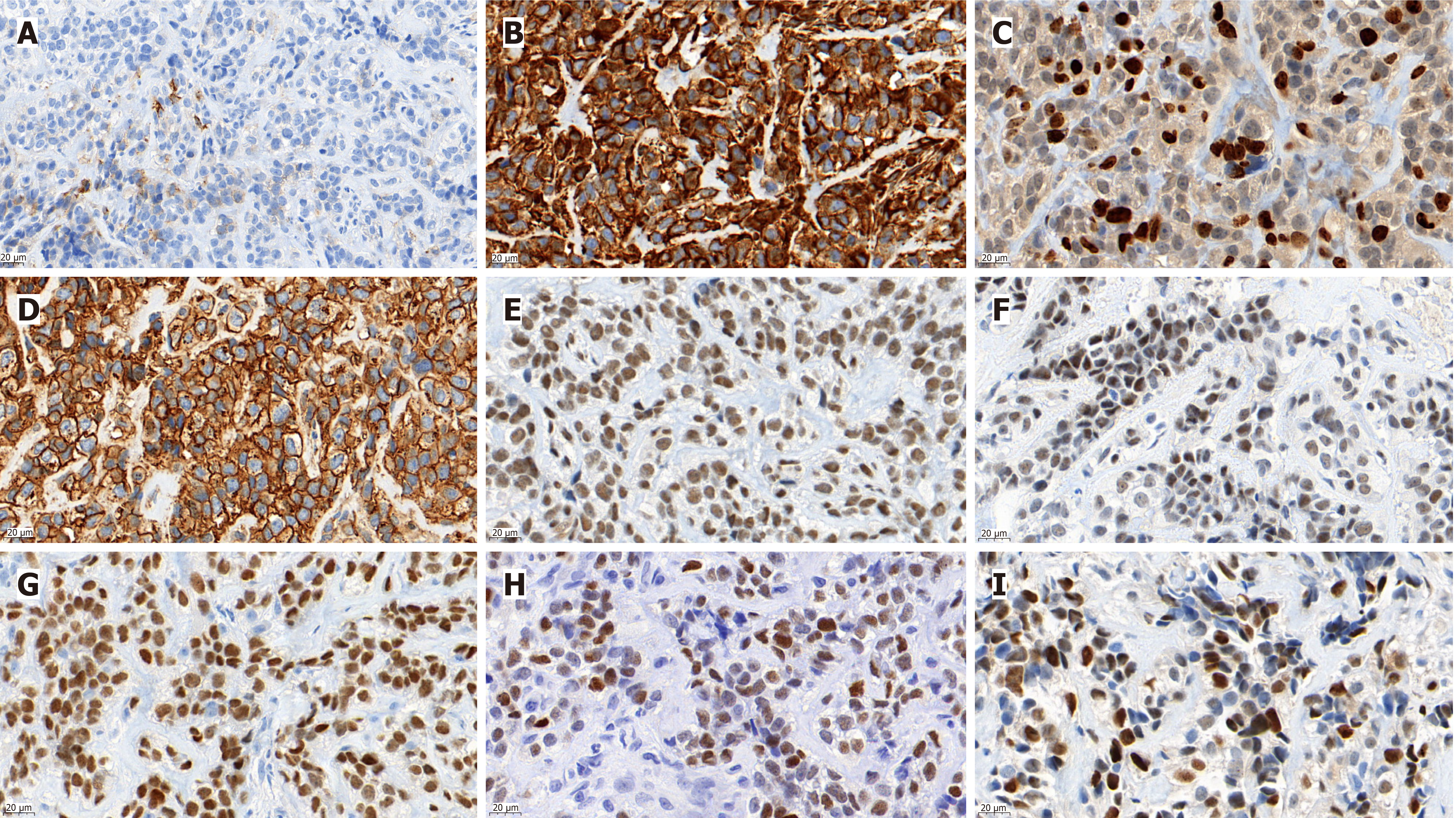
Figure 1 Immunohistochemically, the tumor cells were positive for Vimentin, CD99, NKX2.
2, NKX3.1, INI-1, CyclinD1 and FLI1, and focally positive for epithelial membrane antigen. A: Immunohistochemistry showed a small number of epithelial membrane antigen positives in tumor cells; B: The tumor cells were diffusely positive for Vimentin; C: About 25% of the tumor cells were positive for Ki67; D: Tumor cells were also permeated with CD99 positivity; E-I: Positive for INI1, FLI1, NKX2.2, NKX3.1, and CyclinD1, respectively. Scale bars are in the lower left corner of each image (original magnification, 630 ×).
- Citation: Hu QL, Zeng C. Clinicopathological analysis of EWSR1/FUS::NFATC2 rearranged sarcoma in the left forearm: A case report. World J Clin Cases 2024; 12(16): 2887-2893
- URL: https://www.wjgnet.com/2307-8960/full/v12/i16/2887.htm
- DOI: https://dx.doi.org/10.12998/wjcc.v12.i16.2887


