Copyright
©The Author(s) 2024.
World J Clin Cases. Jun 6, 2024; 12(16): 2862-2868
Published online Jun 6, 2024. doi: 10.12998/wjcc.v12.i16.2862
Published online Jun 6, 2024. doi: 10.12998/wjcc.v12.i16.2862
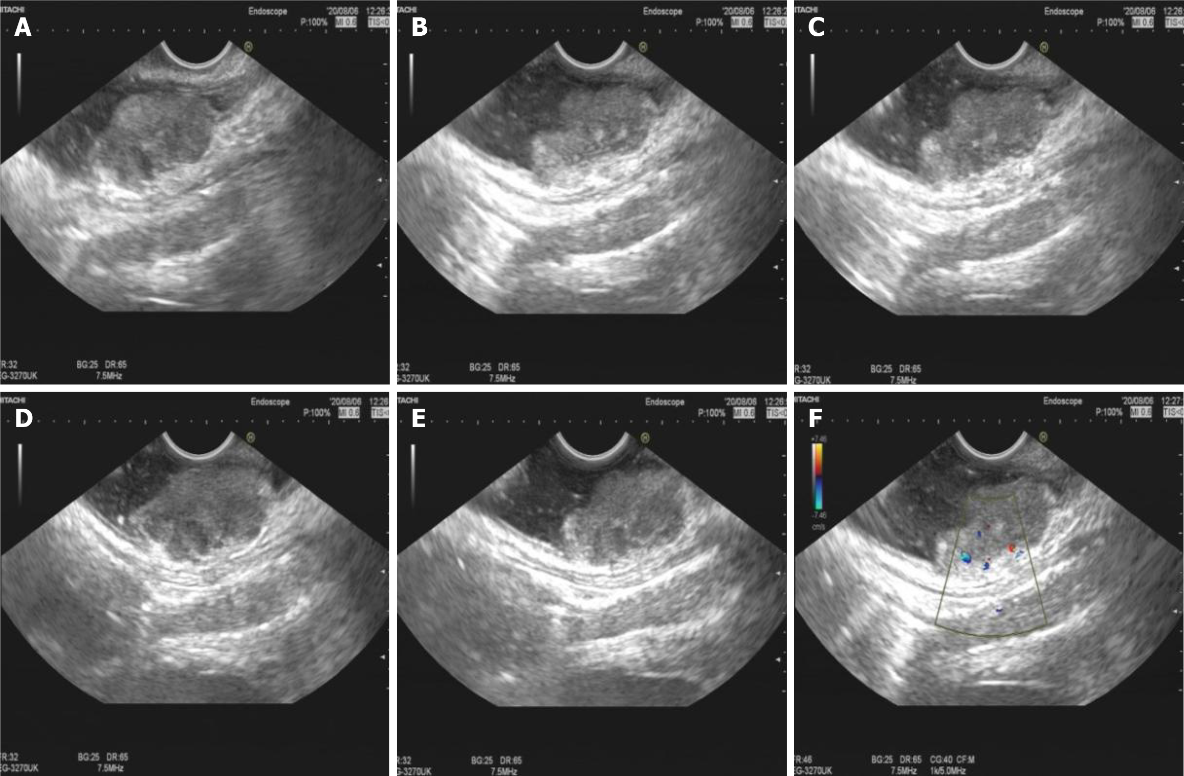
Figure 2 Endoscopic ultrasound image and color Doppler image.
A-E: Endoscopic ultrasound image showing a hypoechoic mucosal mass with involvement of the submucosal layer and heterogeneity of the internal echoes; F: Color Doppler showed localized blood with the intrinsic musculature still intact.
- Citation: Xiong ZE, Wei XX, Wang L, Xia C, Li ZY, Long C, Peng B, Wang T. Endoscopic ultrasound features of rectal melanoma: A case report and review of literature. World J Clin Cases 2024; 12(16): 2862-2868
- URL: https://www.wjgnet.com/2307-8960/full/v12/i16/2862.htm
- DOI: https://dx.doi.org/10.12998/wjcc.v12.i16.2862


