Copyright
©The Author(s) 2024.
World J Clin Cases. Jun 6, 2024; 12(16): 2831-2836
Published online Jun 6, 2024. doi: 10.12998/wjcc.v12.i16.2831
Published online Jun 6, 2024. doi: 10.12998/wjcc.v12.i16.2831
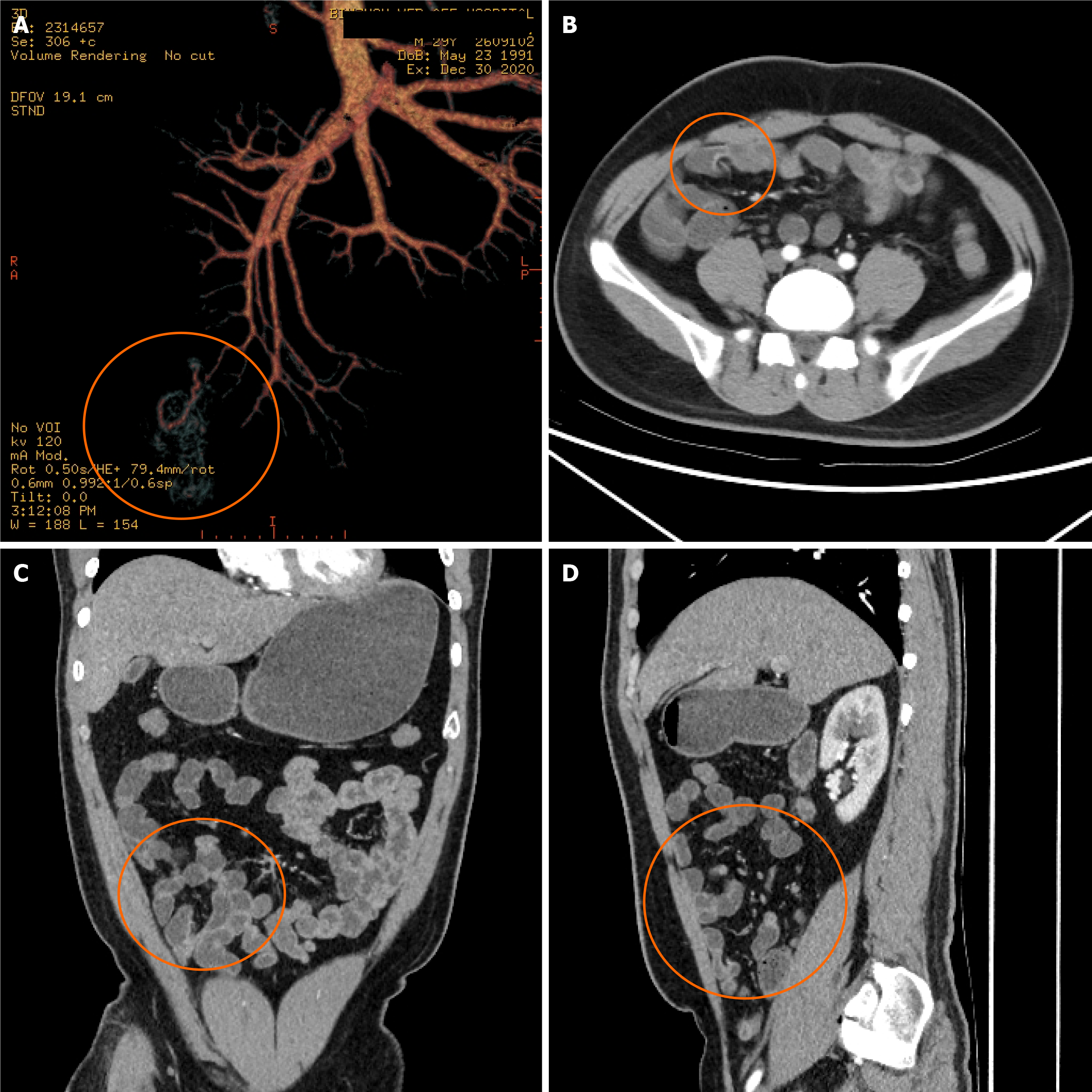
Figure 1 Three-dimensional computed tomography reconstruction of the small intestine, showing the middle part of the ileum.
A-D: The portal vein branches can be seen entering the intestinal wall and the intestinal wall encircles the portal vein branches and corresponding mesenteric adipose tissues from the side and protrudes into the intestinal cavity with a total length of about 10 cm.
- Citation: Zhang SH, Fan MW, Chen Y, Hu YB, Liu CX. Computed tomography three-dimensional reconstruction in the diagnosis of bleeding small intestinal polyps: A case report. World J Clin Cases 2024; 12(16): 2831-2836
- URL: https://www.wjgnet.com/2307-8960/full/v12/i16/2831.htm
- DOI: https://dx.doi.org/10.12998/wjcc.v12.i16.2831


