Copyright
©The Author(s) 2024.
World J Clin Cases. Jun 6, 2024; 12(16): 2822-2830
Published online Jun 6, 2024. doi: 10.12998/wjcc.v12.i16.2822
Published online Jun 6, 2024. doi: 10.12998/wjcc.v12.i16.2822
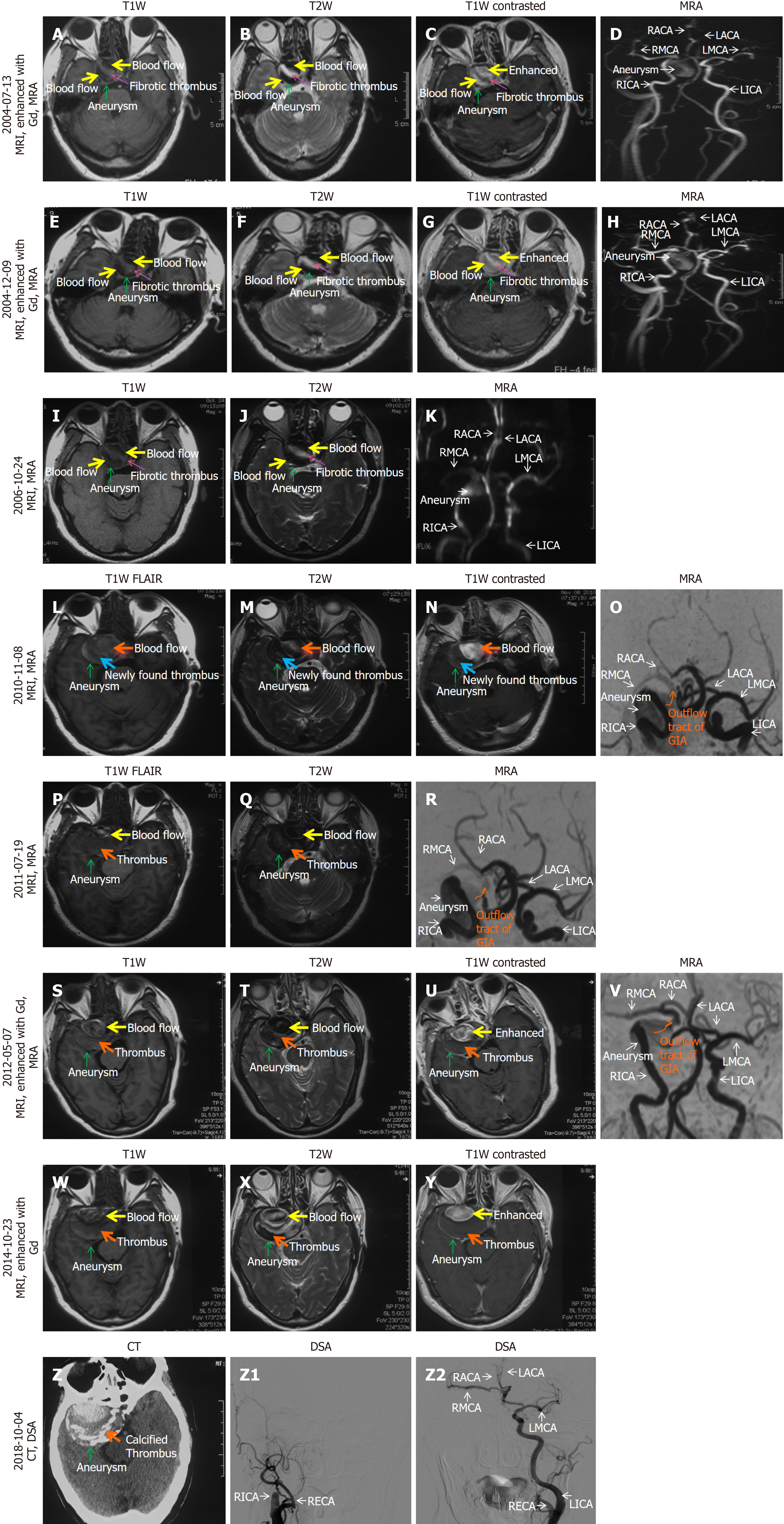
Figure 1 Imaging displaying the sequentially occurred growth of a giant cavernous carotid aneurysm, progression of aneurysmal thrombosis, aneurysmal calcification, spontaneous occlusion of ipsilateral internal carotid artery and closure of aneurysm in our patient.
A-D: On July 13, 2004 [30 mm (axial) × 38 mm (coronal) × 28 mm (sagittal) in diameter], a head magnetic resonance imaging (MRI) (A and B), an enhanced MRI (C) with gadolinium-diethylenetriaminepentaacetic acid and magnetic resonance angiography (MRA) (D) detected a giant intracranial aneurysm (GIA) in the cavernous sinus segment of the right internal carotid artery (RICA). The GIA contained blood flow mainly (yellow arrows on A-C) and fibrotic thrombus (pink arrows on A, B, C) in the center of the GIA, and the RICA was unobstructed (D); E-K: On December 9, 2004 (E-H) [31 mm (axial) × 39 mm (coronal) × 28 mm (sagittal) in diameter] and October 24, 2006 (I-K) [34 mm (axial) × 33 mm (sagittal) in diameter], the MRI and MRA showed that this GIA had not significantly enlarged, the GIA still contained blood flow (yellow arrows on E-G, I, and J) mainly and fibrotic thrombus (pink arrows on E-G, I, and J) in the center of the GIA, and their proportions of components did not change compared to July 13, 2004 (A-C); L-O: On November 8, 2010 [37 mm (axial) × 39 mm (sagittal) in diameter], the MRI showed that the GIA had become enlarged with more newly found thrombus (blue arrows on L-N) than blood flow (orange arrows on L-N). The MRA (O) showed that the signal intensities of right middle cerebral artery (RMCA) and right anterior cerebral artery (RACA) were lower than those on LMCA and left anterior cerebral artery on November 8, 2010, and those on RMCA and RACA on MRA examined on July 13, 2004 (D), December 9, 2004 (H), and October 24, 2006 (K), suggesting a reduced blood supply to the RICA distal to the GIA (RMCA and RACA). The outflow tract of the GIA (red curve on O) and the RICA proximal to the GIA were still visible, indicating that the entire RICA was still unobstructed at this stage; P-Y: Compared to the MRI and MRA examinations on November 8, 2010 (L-O), the examinations on July 19, 2011 (P-R) [39 mm (axial) × 40 mm (coronal) × 39 mm (sagittal) in diameter], May 7, 2012 (S-V) [56 mm (axial) × 52 mm (coronal) × 44 mm (sagittal) in diameter] and October 23, 2014 (W-Y) [56 mm (axial) × 52 mm (coronal) × 46 mm (sagittal) in diameter] showed significant growth of the GIA and an increased proportion of thrombus compared to the blood flow inside the GIA. The outflow tract of the GIA (red curve on image R and V) and the RICA proximal to the GIA were still visible, indicating that the entire RICA was still unobstructed at this stage; Z-z2: On October 4, 2018 [54 mm (axial) in diameter], a CT scan detected a concentric hyperdense (calcified) mass inside the GIA (Z), and DSA showed that the GIA was not visible (z1), indicating complete closure of the GIA, and the RICA proximal and distal to the GIA was completely invisible (z1), meaning that the entire RICA was completely occluded, RMCA and RACA were supplied by LICA via the anterior communicating artery (z2). The structure and three-dimension morphology of the GIA could not be obtained because our patient refused to undergo CT angiography. MRI: Magnetic resonance imaging; MRA: Magnetic resonance angiography; Gd-DTPA: Gadolinium-diethylenetriaminepentaacetic acid; CT: Computed tomography; DSA: Digital subtraction angiography; 3D: Three-dimension; CTA: Computed tomography angiography; GIA: Giant intracranial aneurysm; RICA: Right internal carotid artery; LICA: Left internal carotid artery; RMCA: Right middle cerebral artery; LMCA: Left middle cerebral artery; RACA: Right anterior cerebral artery; LACA: Left anterior cerebral artery; RECA: Right external cerebral artery.
- Citation: Wang MX, Nie QB. Giant cavernous aneurysms occluded by aneurysmal thrombosis, calcification, parent artery occlusion: A case report and review of literature. World J Clin Cases 2024; 12(16): 2822-2830
- URL: https://www.wjgnet.com/2307-8960/full/v12/i16/2822.htm
- DOI: https://dx.doi.org/10.12998/wjcc.v12.i16.2822


