Copyright
©The Author(s) 2024.
World J Clin Cases. Jun 6, 2024; 12(16): 2722-2728
Published online Jun 6, 2024. doi: 10.12998/wjcc.v12.i16.2722
Published online Jun 6, 2024. doi: 10.12998/wjcc.v12.i16.2722
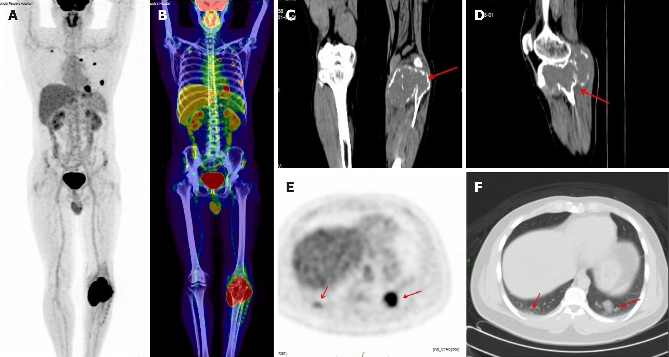
Figure 2 Left upper tibia giant cell tumor of bone invading the upper fibula with multiple lung metastases on positron emission tomo
- Citation: Kou MQ, Xu BQ, Liu HT. Multimodal imaging in the diagnosis of bone giant cell tumors: A retrospective study. World J Clin Cases 2024; 12(16): 2722-2728
- URL: https://www.wjgnet.com/2307-8960/full/v12/i16/2722.htm
- DOI: https://dx.doi.org/10.12998/wjcc.v12.i16.2722


