Copyright
©The Author(s) 2024.
World J Clin Cases. May 26, 2024; 12(15): 2678-2681
Published online May 26, 2024. doi: 10.12998/wjcc.v12.i15.2678
Published online May 26, 2024. doi: 10.12998/wjcc.v12.i15.2678
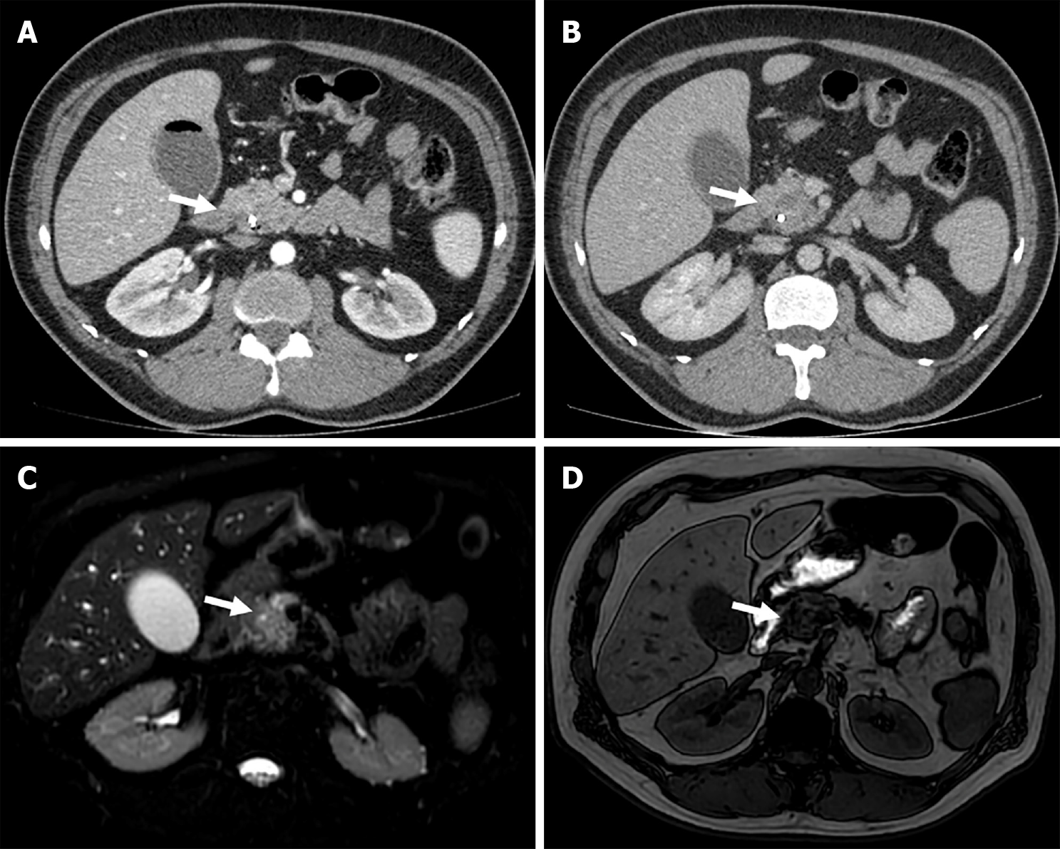
Figure 3 Pancreatic ductal adenocarcinoma in a 72-years-old man with obstructive jaundice.
A: Axial contrast-enhanced computed tomography image shows a well-circumscribed exophytic tumor in the pancreatic head with heterogeneuos enhancement in delayed arterial; B: Portovenous phase; C: Axial magnetic resonance fat suppression T2-weighted images image showing a mildly hyperdense irregular mass in the head of the pancreas; D: T1-weighted images with hypointense signal.
- Citation: Lindner C. Imaging features of malignant vs stone-induced biliary obstruction: Aspects to consider. World J Clin Cases 2024; 12(15): 2678-2681
- URL: https://www.wjgnet.com/2307-8960/full/v12/i15/2678.htm
- DOI: https://dx.doi.org/10.12998/wjcc.v12.i15.2678


