Copyright
©The Author(s) 2024.
World J Clin Cases. May 26, 2024; 12(15): 2672-2677
Published online May 26, 2024. doi: 10.12998/wjcc.v12.i15.2672
Published online May 26, 2024. doi: 10.12998/wjcc.v12.i15.2672
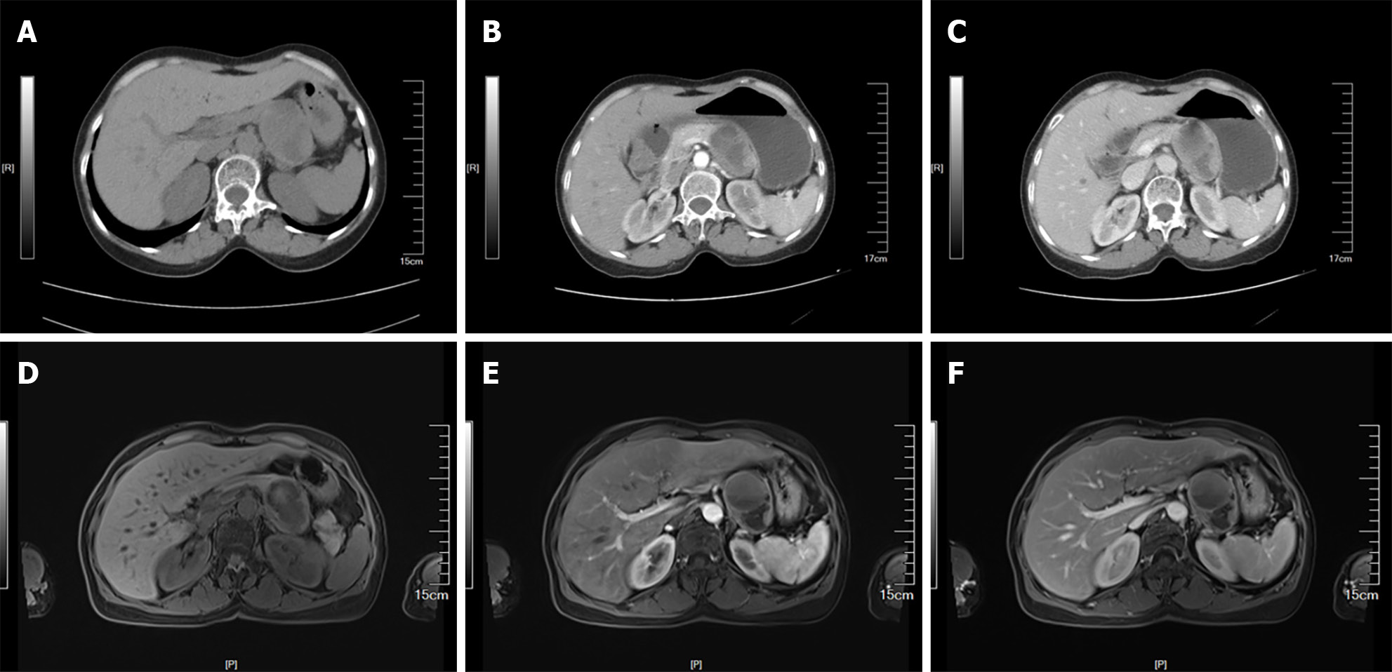
Figure 1 Abdominal computed tomography and magnetic resonance imaging show a 6.
5 cm × 4.5 cm cystic solid mass in the left upper abdomen with heterogeneous density and mild enhancement in the arterial phase. A: Computed tomography (CT) plain scan period; B: CT arterial phase; C: CT venous phase; D: Magnetic resonance imaging (MRI) plain scan period; E: MRI arterial phase; F: MRI venous phase.
- Citation: Kang LM, Yu FK, Zhang FW, Xu L. Subclinical paraganglioma of the retroperitoneum: A case report. World J Clin Cases 2024; 12(15): 2672-2677
- URL: https://www.wjgnet.com/2307-8960/full/v12/i15/2672.htm
- DOI: https://dx.doi.org/10.12998/wjcc.v12.i15.2672


