Copyright
©The Author(s) 2024.
World J Clin Cases. May 26, 2024; 12(15): 2655-2663
Published online May 26, 2024. doi: 10.12998/wjcc.v12.i15.2655
Published online May 26, 2024. doi: 10.12998/wjcc.v12.i15.2655
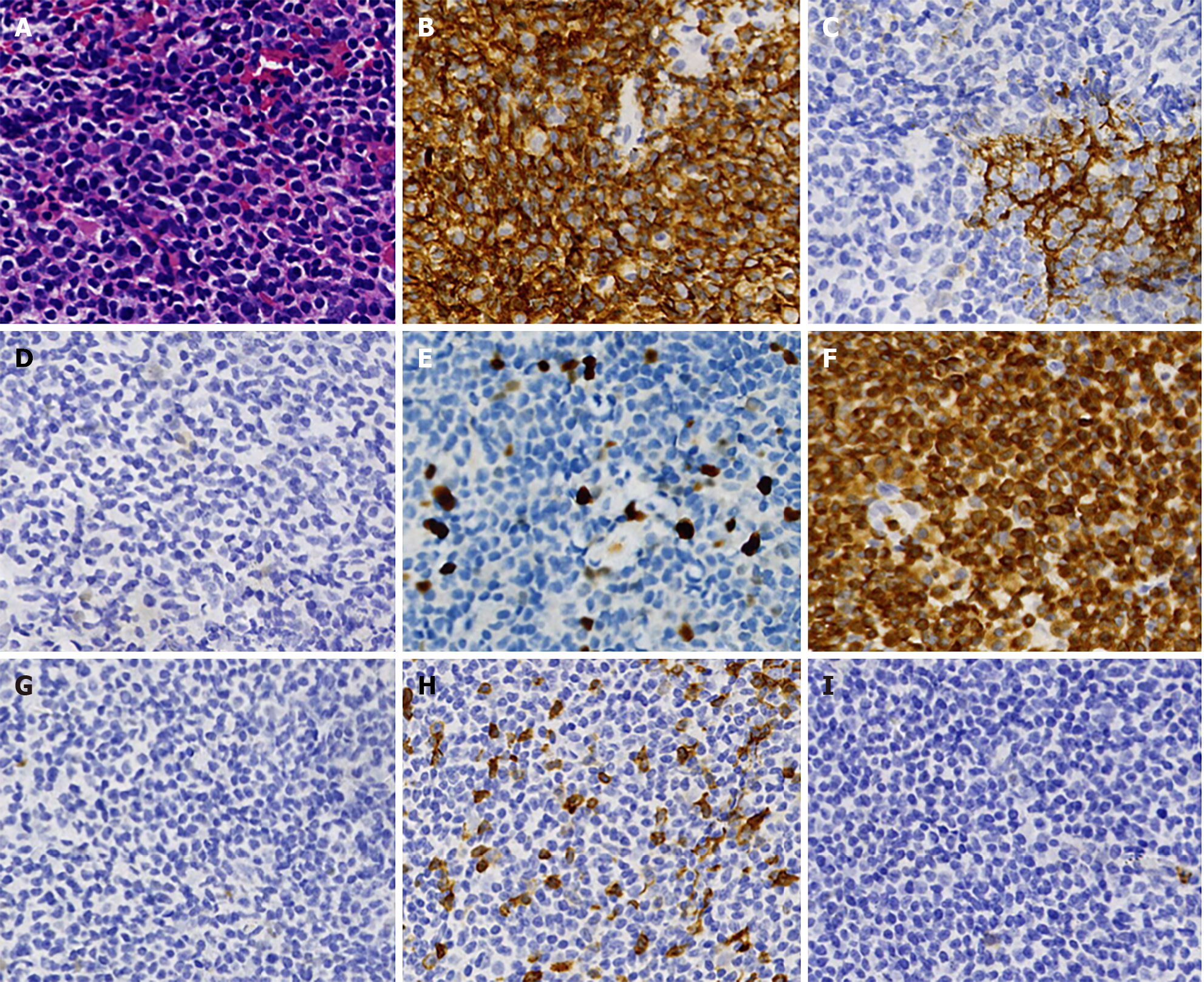
Figure 1 Immunohistochemistry and cellular morphology of the first lymph node biopsy.
A: Tumor cells are diffuse, small to medium in size, with a slightly irregular nucleus. The lymphocytic hyperplasia shows partial cytoplasmic empty and a fuzzy nodular distribution (hematoxylin and eosin, 400 ×); B-I: The majority of the infiltrating cells were positive for CD20 (B), PAX5, B-cell lymphoma-2 (BCL-2) (F), CD21, CD23 (C), and negative for CD10 (I), CD5 (H), BCL-6 (G), and CyclinD1 (D). Their Ki-67 (E) showed approximately 10% (EnVision, 400 ×).
- Citation: Fan ZM, Wu DL, Xu NW, Ye L, Yan LP, Li LJ, Zhang JY. Transformation of marginal zone lymphoma into high-grade B-cell lymphoma expressing terminal deoxynucleotidyl transferase: A case report. World J Clin Cases 2024; 12(15): 2655-2663
- URL: https://www.wjgnet.com/2307-8960/full/v12/i15/2655.htm
- DOI: https://dx.doi.org/10.12998/wjcc.v12.i15.2655


