Copyright
©The Author(s) 2024.
World J Clin Cases. May 26, 2024; 12(15): 2627-2635
Published online May 26, 2024. doi: 10.12998/wjcc.v12.i15.2627
Published online May 26, 2024. doi: 10.12998/wjcc.v12.i15.2627
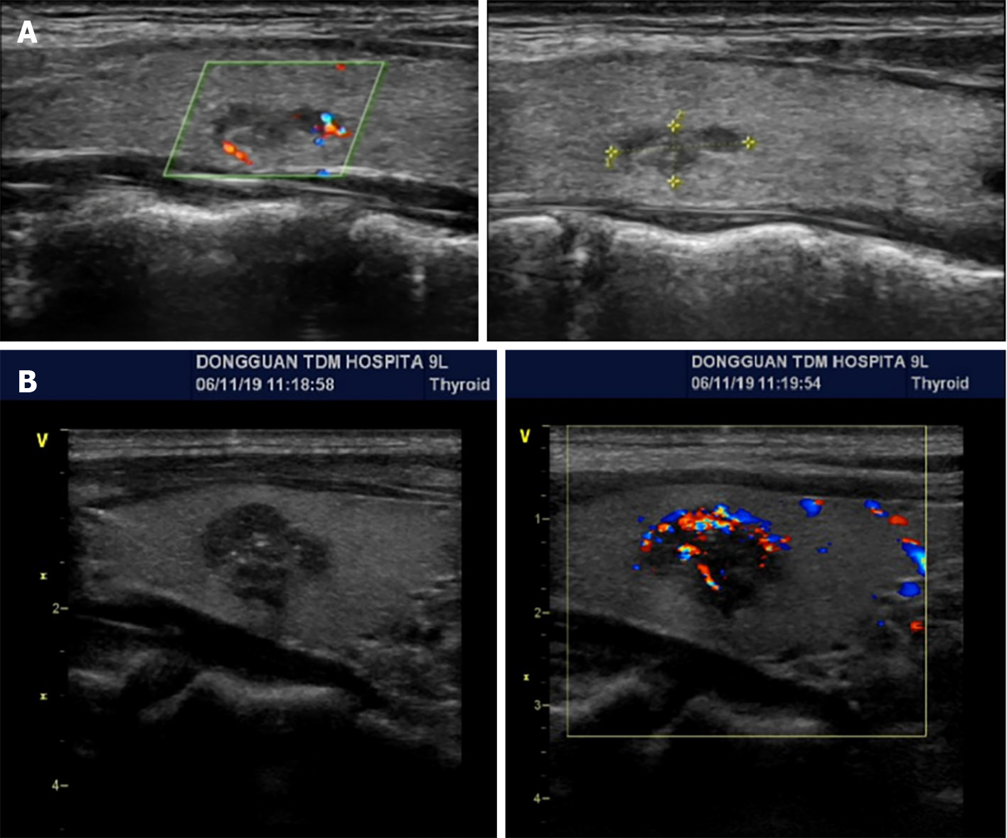
Figure 2 Thyroid B-ultrasound.
A: Proband IIa. Bilateral thyroid nodules were observed (10 mm × 4 mm); B: Patient Ib. A hypoechoic nodule (16 mm × 11 mm) was observed in the right thyroid.
- Citation: Zhang HF, Huang SL, Wang WL, Zhou YQ, Jiang J, Dai ZJ. C634Y mutation in RET-induced multiple endocrine neoplasia type 2A: A case report. World J Clin Cases 2024; 12(15): 2627-2635
- URL: https://www.wjgnet.com/2307-8960/full/v12/i15/2627.htm
- DOI: https://dx.doi.org/10.12998/wjcc.v12.i15.2627


