Copyright
©The Author(s) 2024.
World J Clin Cases. May 26, 2024; 12(15): 2621-2626
Published online May 26, 2024. doi: 10.12998/wjcc.v12.i15.2621
Published online May 26, 2024. doi: 10.12998/wjcc.v12.i15.2621
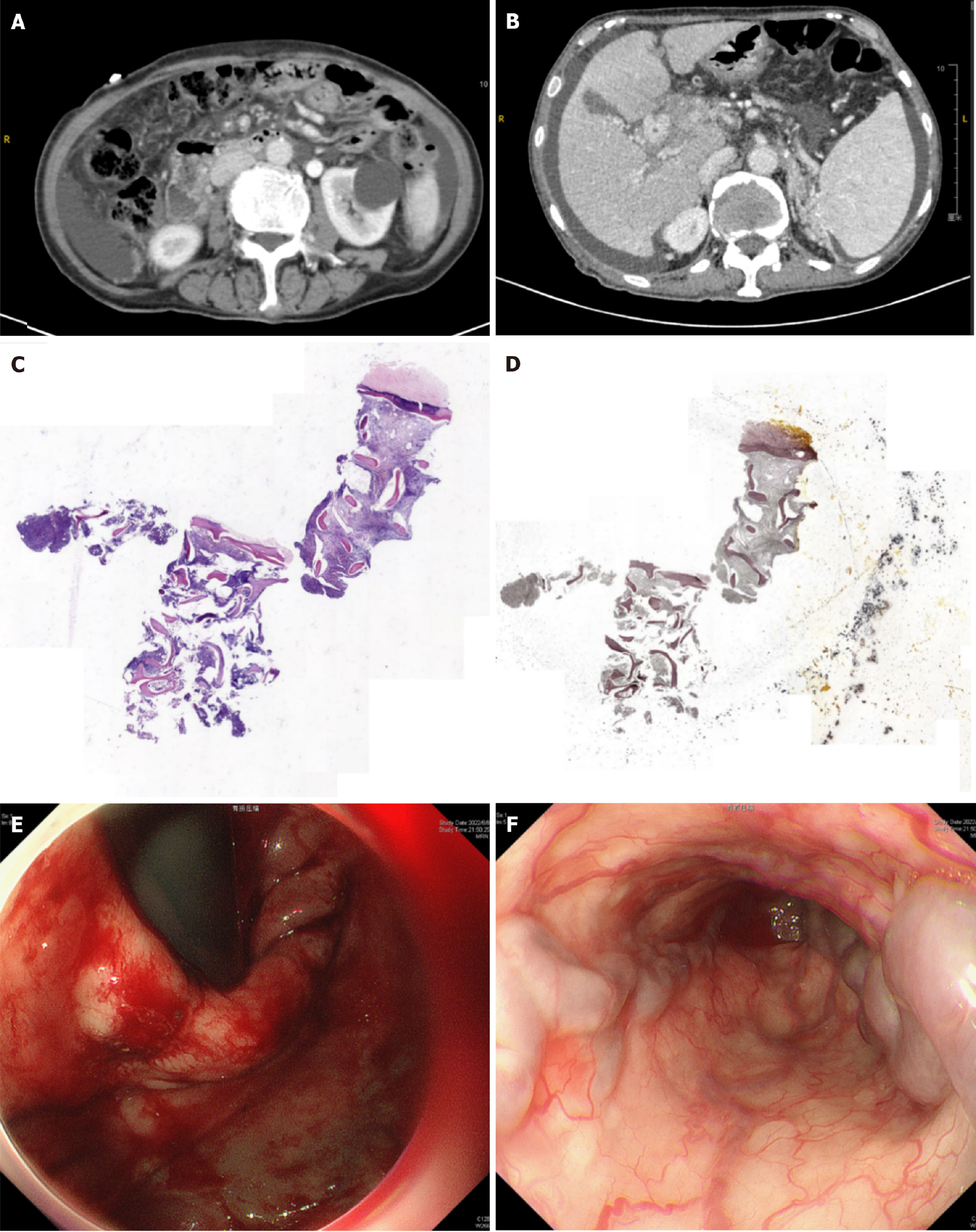
Figure 1 Examinations images.
A and B: Contrast-enhanced abdominal computerized tomography image demonstrating extensive splanchnic vein thrombosis involving the portal, splenic, and superior mesenteric veins; C amd D: Bone marrow biopsy revealed that myelofibrosis (MF-2 grade) was accompanied by a significant increase in platelet count; E and F: An emergency endoscopy showed esophagogastric varices (F3CbRc+Les, severe). A white thrombus was detected on the middle of the esophageal varices.
- Citation: Chen Y, Kong BB, Yin H, Liu H, Wu S, Xu T. Acute upper gastrointestinal bleeding due to portal hypertension in a patient with primary myelofibrosis: A case report. World J Clin Cases 2024; 12(15): 2621-2626
- URL: https://www.wjgnet.com/2307-8960/full/v12/i15/2621.htm
- DOI: https://dx.doi.org/10.12998/wjcc.v12.i15.2621


