Copyright
©The Author(s) 2024.
World J Clin Cases. May 26, 2024; 12(15): 2606-2613
Published online May 26, 2024. doi: 10.12998/wjcc.v12.i15.2606
Published online May 26, 2024. doi: 10.12998/wjcc.v12.i15.2606
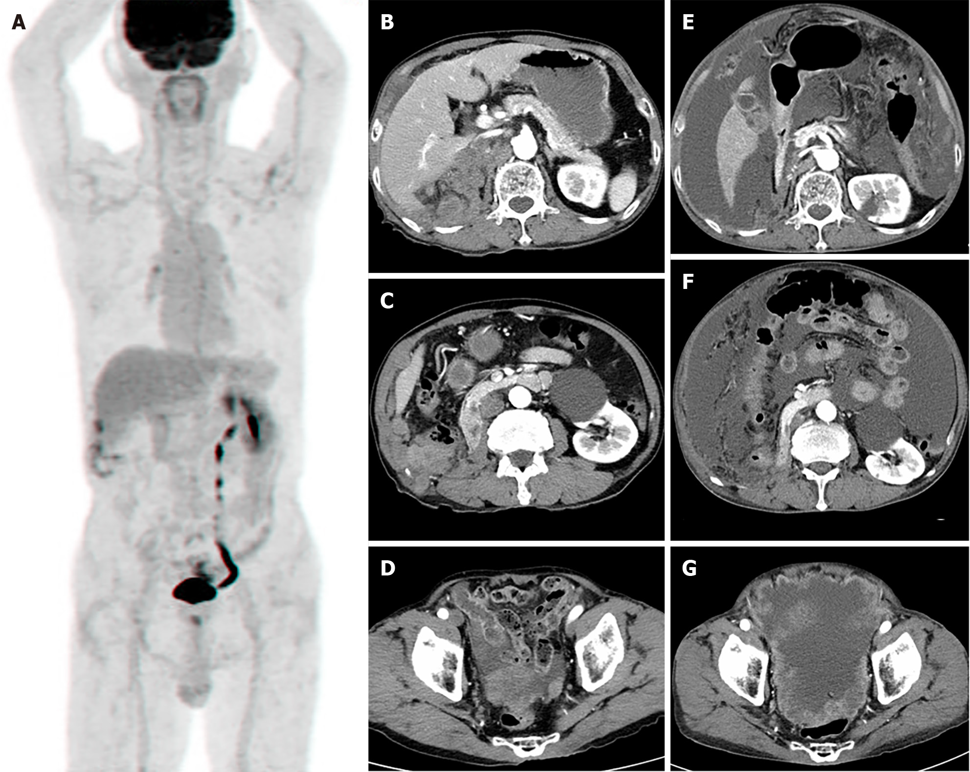
Figure 3 Immediate post-operative 18F-fluorodeoxyglucose torso positron emission tomography scan and follow up abdominal computed tomography.
A: 18F-fluorodeoxyglucose (FDG) torso positron emission tomography after right nephrectomy shows mild FDG uptake at operation site, which suggested postoperative changes. No other abnormal FDG uptake was demonstrated; B-D: Abdominal computed tomography (CT), taken 6 months after right nephrectomy, shows local recurrence and peritoneal metastasis; E-G: Abdominal CT scan, taken 8 months after right nephrectomy, shows locally controlled local recurrence result from radiotherapy but more progression of peritoneal metastasis with increased malignant ascites.
- Citation: Kim S, Park J, Ko YH, Kwon HJ. Primary Ewing sarcoma of the kidney mimicking cystic papillary renal cell carcinoma in an older patient: A case report. World J Clin Cases 2024; 12(15): 2606-2613
- URL: https://www.wjgnet.com/2307-8960/full/v12/i15/2606.htm
- DOI: https://dx.doi.org/10.12998/wjcc.v12.i15.2606


