Copyright
©The Author(s) 2024.
World J Clin Cases. May 26, 2024; 12(15): 2597-2605
Published online May 26, 2024. doi: 10.12998/wjcc.v12.i15.2597
Published online May 26, 2024. doi: 10.12998/wjcc.v12.i15.2597
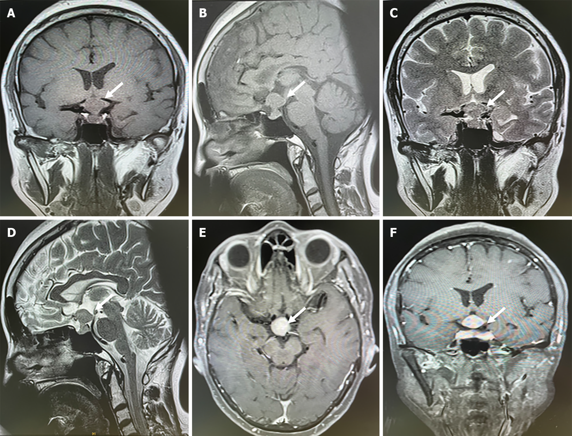
Figure 1 Sellar magnetic resonance imaging showed a sellar lesion related to carotid artery.
A-D: The leision located in the sellar region presented with an isointense signal on T1 and T2-weighted images and could see hourglass sign; E and F: The leision was intensified homogenously enhanced after contrast magnetic resonance imaging.
- Citation: Wang Q, Liu XW, Chen KY. Pituitary metastasis from lung adenocarcinoma: A case report. World J Clin Cases 2024; 12(15): 2597-2605
- URL: https://www.wjgnet.com/2307-8960/full/v12/i15/2597.htm
- DOI: https://dx.doi.org/10.12998/wjcc.v12.i15.2597


