Copyright
©The Author(s) 2024.
World J Clin Cases. May 16, 2024; 12(14): 2451-2456
Published online May 16, 2024. doi: 10.12998/wjcc.v12.i14.2451
Published online May 16, 2024. doi: 10.12998/wjcc.v12.i14.2451
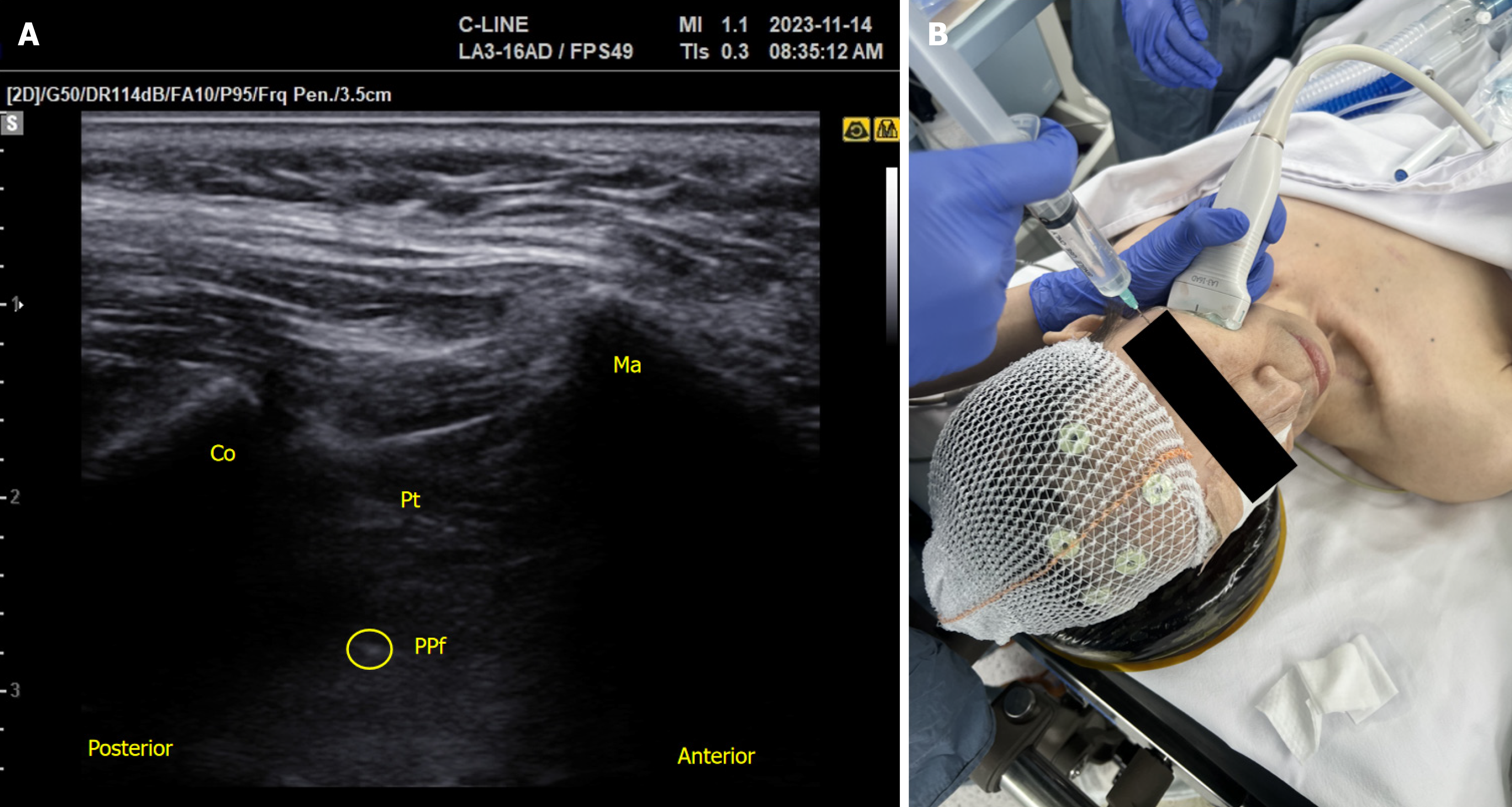
Figure 2 Sphenopalatine ganglion block guided by ultrasonography.
A: Ultrasound image visualizing the surrounding anatomical structures; B: Ultrasound-guided needle placement to perform the sphenopalatine ganglion block. Co: Coronoid process of the mandible; Ma: Maxilla; Pt: Pterygoid muscles; PPf: Pterygopalatine fossa; Yellow circle: Needle tip.
- Citation: Kang H, Park S, Jin Y. Ultrasound-guided sphenopalatine ganglion block for effective analgesia during awake fiberoptic nasotracheal intubation: A case report. World J Clin Cases 2024; 12(14): 2451-2456
- URL: https://www.wjgnet.com/2307-8960/full/v12/i14/2451.htm
- DOI: https://dx.doi.org/10.12998/wjcc.v12.i14.2451


