Copyright
©The Author(s) 2024.
World J Clin Cases. May 16, 2024; 12(14): 2396-2403
Published online May 16, 2024. doi: 10.12998/wjcc.v12.i14.2396
Published online May 16, 2024. doi: 10.12998/wjcc.v12.i14.2396
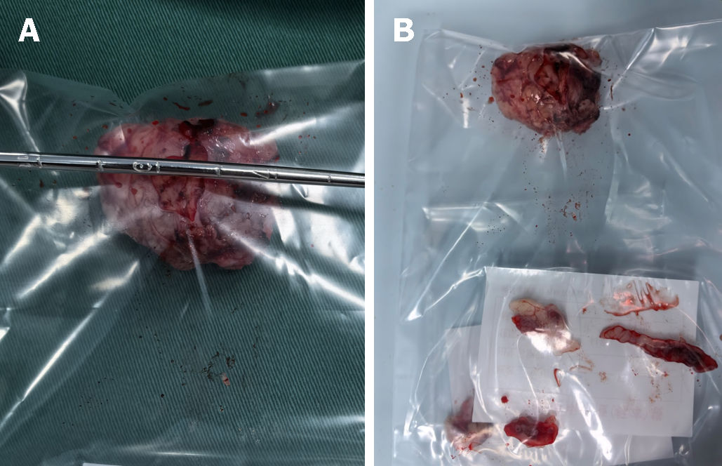
Figure 2 Gross pathological picture of the patient’s vaginal pleomorphic rhabdomyosarcoma.
A: The size of the vaginal mass was approximately 32 mm × 30 mm × 30 mm; B: Gross pathological picture of the vaginal mass, which had no obvious capsule, was pale red with a medium texture, and resembled a uterine leiomyoma. Below, is the 1 cm portion of the vaginal wall that was adjacent to the tumor.
- Citation: Xu P, Ling SS, Hu E, Yi BX. Pleomorphic rhabdomyosarcoma of the vagina: A case report. World J Clin Cases 2024; 12(14): 2396-2403
- URL: https://www.wjgnet.com/2307-8960/full/v12/i14/2396.htm
- DOI: https://dx.doi.org/10.12998/wjcc.v12.i14.2396


