Copyright
©The Author(s) 2024.
World J Clin Cases. May 6, 2024; 12(13): 2254-2262
Published online May 6, 2024. doi: 10.12998/wjcc.v12.i13.2254
Published online May 6, 2024. doi: 10.12998/wjcc.v12.i13.2254
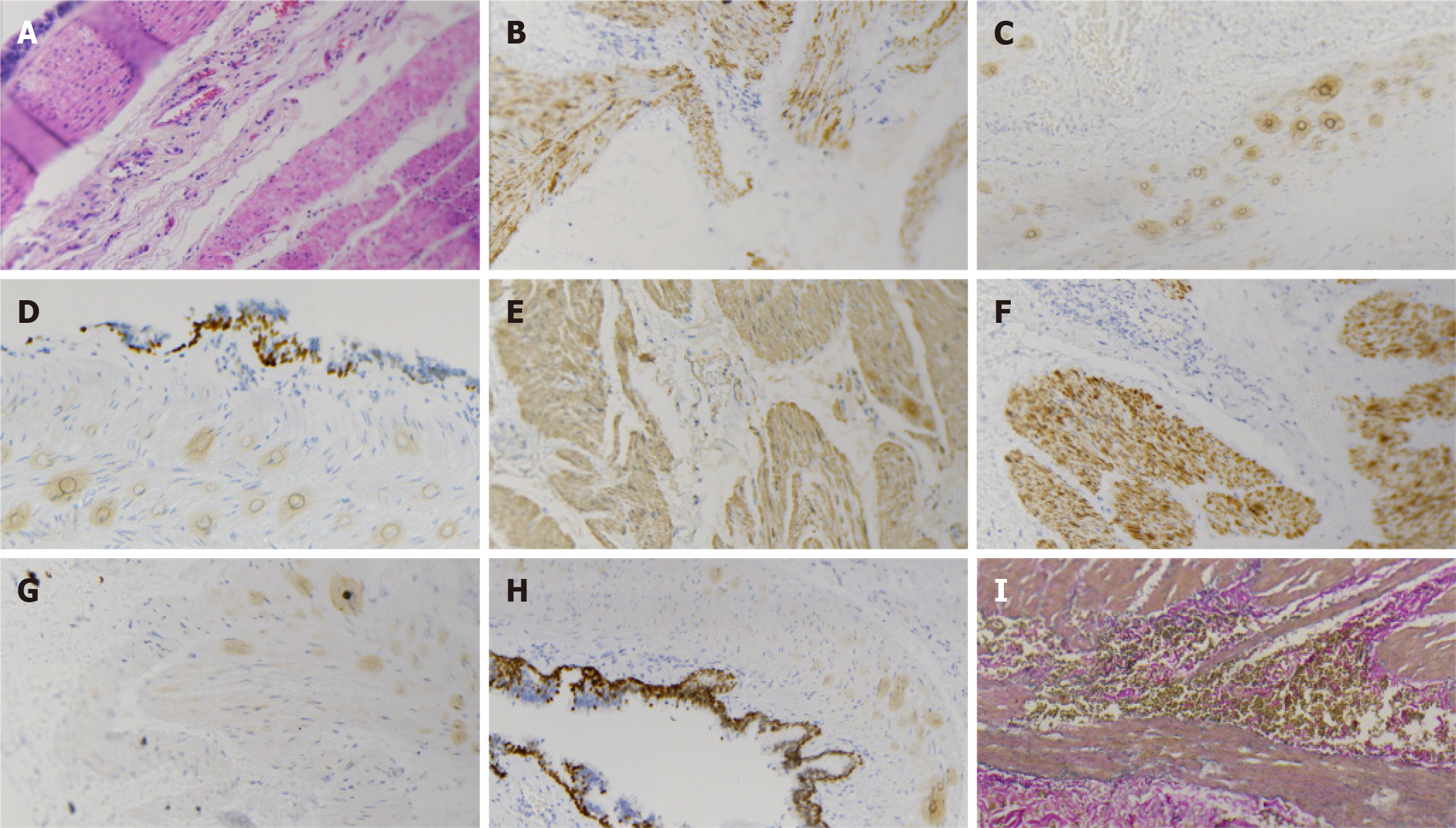
Figure 3 Histopathological analysis and immunohistochemical examination of the resected specimen.
A: Microscopically, pseudostratified ciliated columnar epithelial cells could be observed in the cyst lining, and smooth muscle and small salivary gland tissue could be observed in the cyst; B: Napsin A (+); C: CK20 (-); D: P63 (+); E: SMA (+); F: Desmin (+); G: CK7 (+); H: TTF-1 (+); I: Elastic fiber (+).
- Citation: Lu XR, Jiao XG, Sun QH, Li BW, Zhu QS, Zhu GX, Qu JJ. Young patient with a giant gastric bronchogenic cyst: A case report and review of literature. World J Clin Cases 2024; 12(13): 2254-2262
- URL: https://www.wjgnet.com/2307-8960/full/v12/i13/2254.htm
- DOI: https://dx.doi.org/10.12998/wjcc.v12.i13.2254


