Copyright
©The Author(s) 2024.
World J Clin Cases. May 6, 2024; 12(13): 2231-2236
Published online May 6, 2024. doi: 10.12998/wjcc.v12.i13.2231
Published online May 6, 2024. doi: 10.12998/wjcc.v12.i13.2231
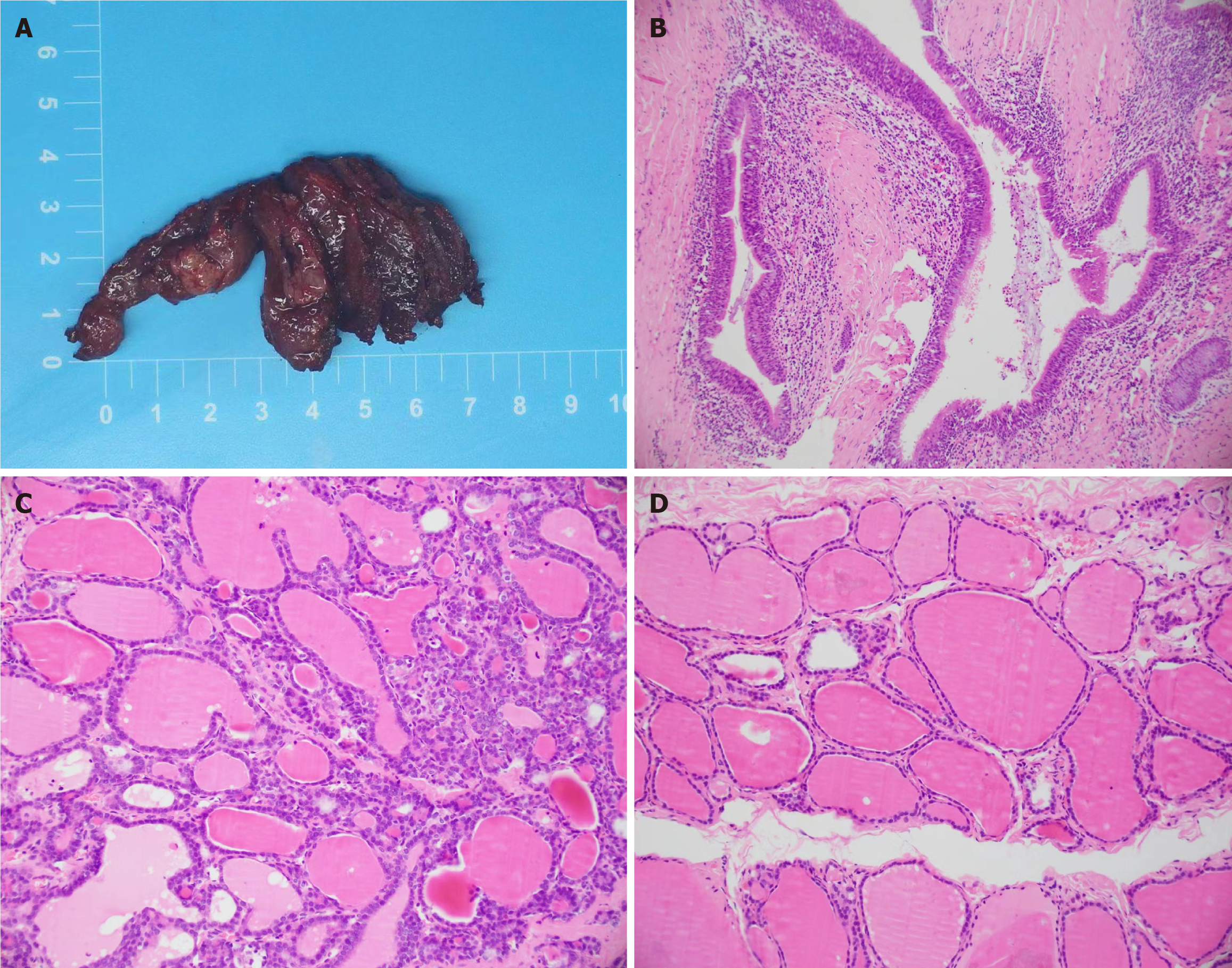
Figure 5 Histopathological analysis of the resected specimen.
A: The resected specimen: Cyst in the left thyroid gland; B-D: Haematoxylin and eosin staining, the cyst wall is ciliated columnar epithelial (× 100).
- Citation: Lin HG, Liu M, Huang XY, Liu DS. Intra-thyroid esophageal duplication cyst: A case report. World J Clin Cases 2024; 12(13): 2231-2236
- URL: https://www.wjgnet.com/2307-8960/full/v12/i13/2231.htm
- DOI: https://dx.doi.org/10.12998/wjcc.v12.i13.2231


