Copyright
©The Author(s) 2024.
World J Clin Cases. Apr 26, 2024; 12(12): 2122-2127
Published online Apr 26, 2024. doi: 10.12998/wjcc.v12.i12.2122
Published online Apr 26, 2024. doi: 10.12998/wjcc.v12.i12.2122
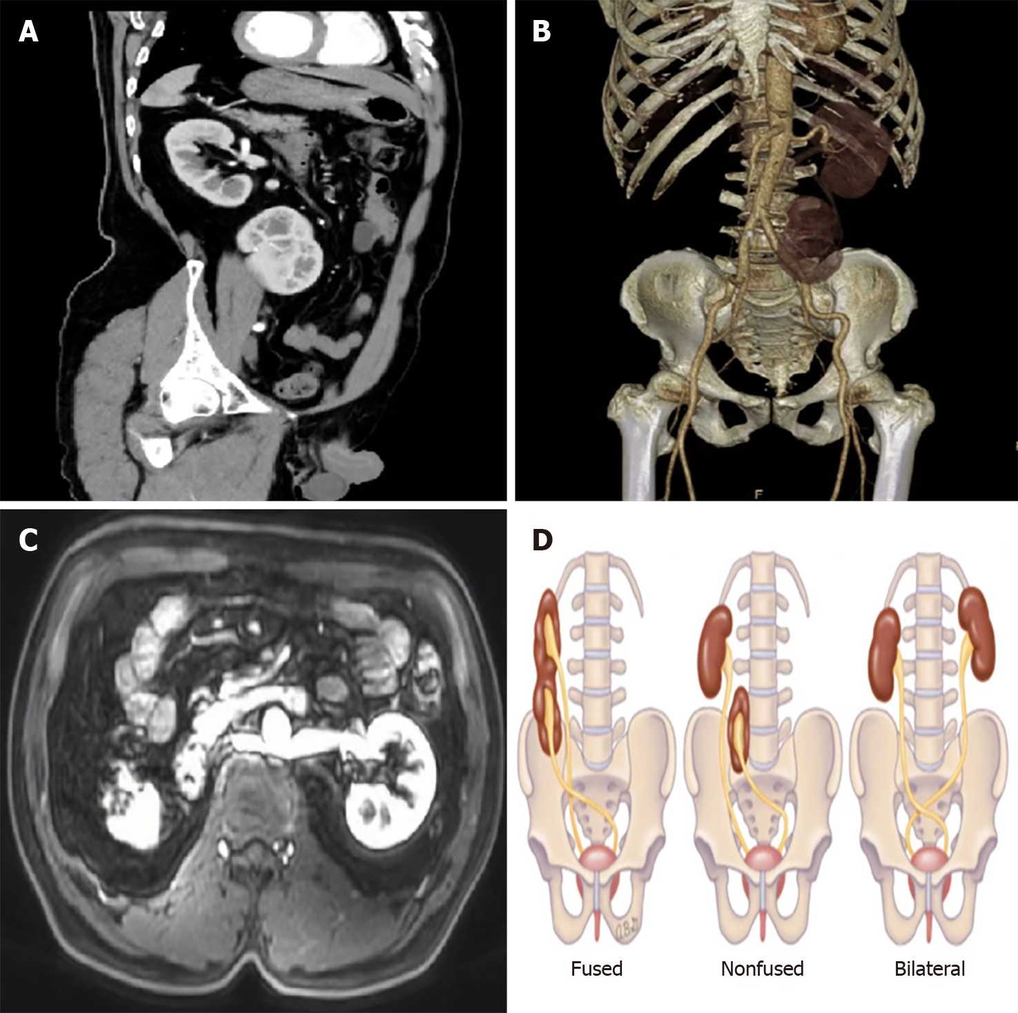
Figure 2 Imaging examination and example images of crossed renal ectopia.
A: Abdominal contrast-enhanced computed tomography Indicating crossed renal ectopia (CRE); B: Three dimensional imaging shows both kidneys on the left side; C: The left renal vein is located behind the abdominal aorta; D: Schematic diagram of CRE.
- Citation: Tang ZW, Yang HF, Wu ZY, Wang CY. Crossed renal ectopia with rectal cancer: A case report. World J Clin Cases 2024; 12(12): 2122-2127
- URL: https://www.wjgnet.com/2307-8960/full/v12/i12/2122.htm
- DOI: https://dx.doi.org/10.12998/wjcc.v12.i12.2122


