Copyright
©The Author(s) 2024.
World J Clin Cases. Apr 26, 2024; 12(12): 2116-2121
Published online Apr 26, 2024. doi: 10.12998/wjcc.v12.i12.2116
Published online Apr 26, 2024. doi: 10.12998/wjcc.v12.i12.2116
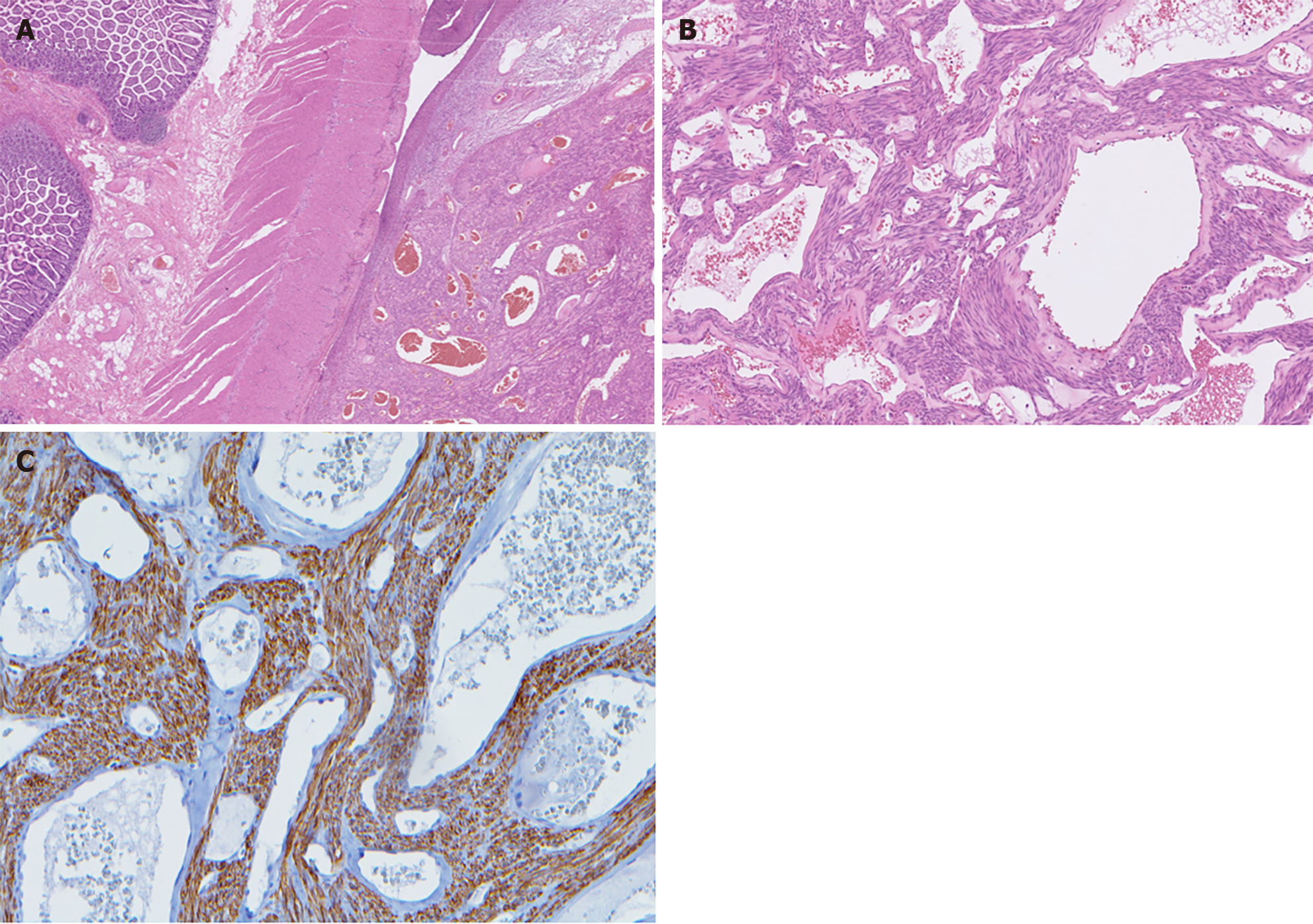
Figure 3 Histopathology of angioleiomyoma.
A: Ileal tissue with a well-circumscribed subserosal tumor; B: Proliferative spindle smooth muscle cells bearing brightly eosinophilic cytoplasm and arranged in fascicles, punctuated by variable-sized vascular channels. The vessels were irregularly dilated with attenuated walls; C: The proliferative smooth muscle cells were highlighted by desmin immunostaining, while the cavernous vascular channels lacked a well-formed muscular wall.
- Citation: Hou TY, Tzeng WJ, Lee PH. Small intestine angioleiomyoma as a rare cause of perforation: A case report. World J Clin Cases 2024; 12(12): 2116-2121
- URL: https://www.wjgnet.com/2307-8960/full/v12/i12/2116.htm
- DOI: https://dx.doi.org/10.12998/wjcc.v12.i12.2116


