Copyright
©The Author(s) 2024.
World J Clin Cases. Apr 26, 2024; 12(12): 2109-2115
Published online Apr 26, 2024. doi: 10.12998/wjcc.v12.i12.2109
Published online Apr 26, 2024. doi: 10.12998/wjcc.v12.i12.2109
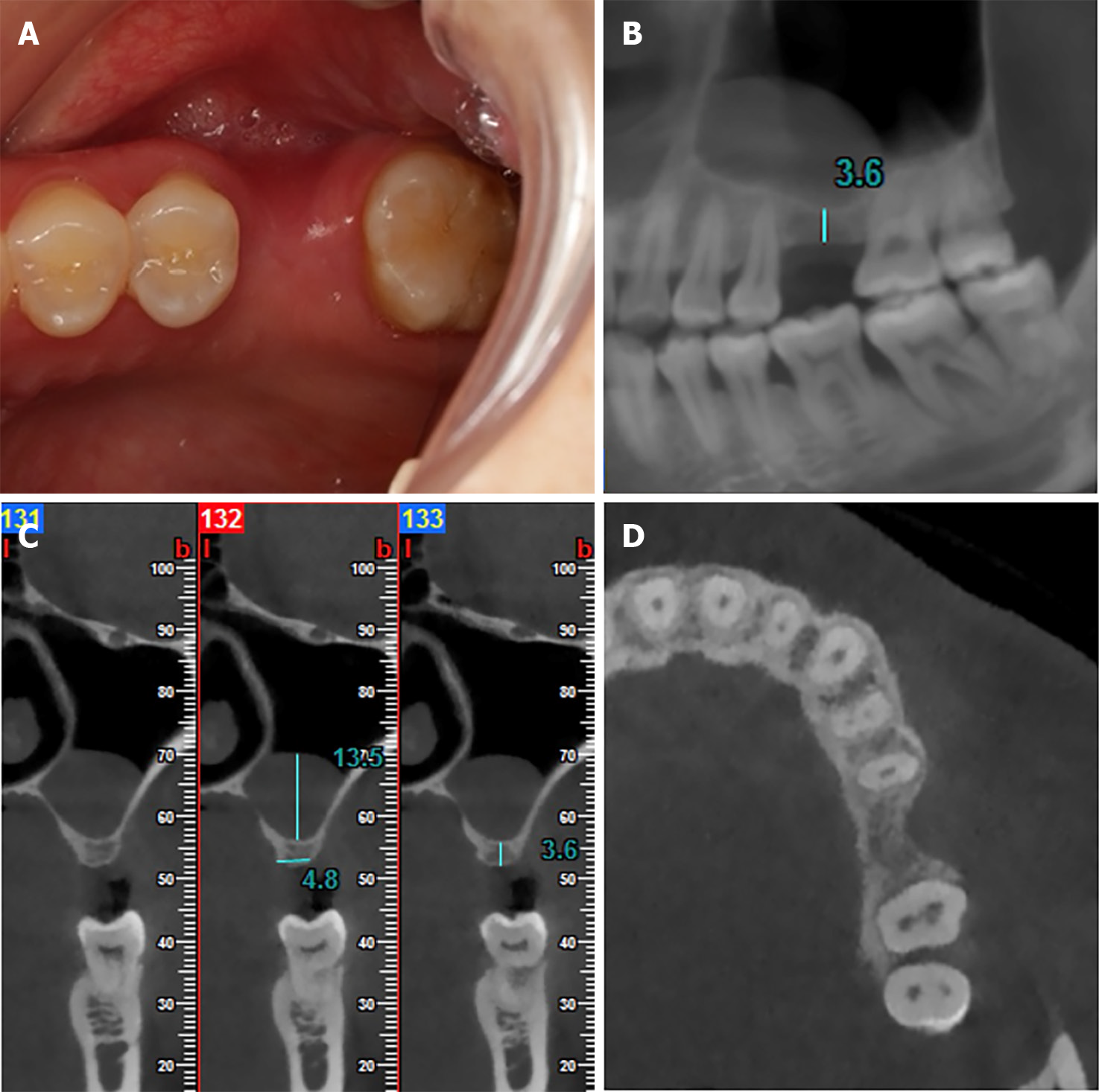
Figure 1 Preoperative occlusal view and cone beam computed tomography images of tooth 26.
A: Occlusal view of tooth 26 with the horizontal bone deficiency; B: Coronal cone beam computed tomography (CBCT) image showed the mesio-distal dimensions and the region of pseudocyst; C: Sagittal CBCT image showed the available bone heights of tooth 26 and the height of pseudocyst; D: Horizontal CBCT image showed horizontal bone deficiency of tooth 26.
- Citation: Wang YL, Shao WJ, Wang M. Bone block from lateral window - correcting vertical and horizontal bone deficiency in maxilla posterior site: A case report. World J Clin Cases 2024; 12(12): 2109-2115
- URL: https://www.wjgnet.com/2307-8960/full/v12/i12/2109.htm
- DOI: https://dx.doi.org/10.12998/wjcc.v12.i12.2109


