Copyright
©The Author(s) 2024.
World J Clin Cases. Apr 26, 2024; 12(12): 2086-2091
Published online Apr 26, 2024. doi: 10.12998/wjcc.v12.i12.2086
Published online Apr 26, 2024. doi: 10.12998/wjcc.v12.i12.2086
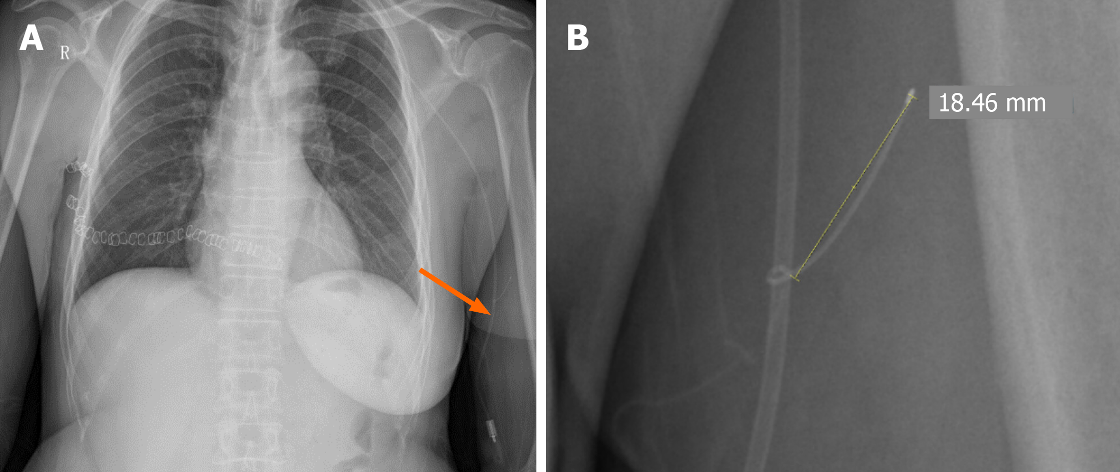
Figure 1 X-ray imaging results.
A: The postoperative X-ray images indicated that the peripherally inserted central catheter was positioned correctly at the T7 cone space level on the right side; B: The postoperative X-ray images revealed that an unusual guide wire measuring around 1.8 cm in length was detectable above the left elbow.
- Citation: Hu CD, Lv R, Zhao YX, Zhang MH, Zeng HD, Mao YW. Basilic vein variation encountered during surgery for arm vein port: A case report. World J Clin Cases 2024; 12(12): 2086-2091
- URL: https://www.wjgnet.com/2307-8960/full/v12/i12/2086.htm
- DOI: https://dx.doi.org/10.12998/wjcc.v12.i12.2086


