Copyright
©The Author(s) 2024.
World J Clin Cases. Apr 16, 2024; 12(11): 1980-1989
Published online Apr 16, 2024. doi: 10.12998/wjcc.v12.i11.1980
Published online Apr 16, 2024. doi: 10.12998/wjcc.v12.i11.1980
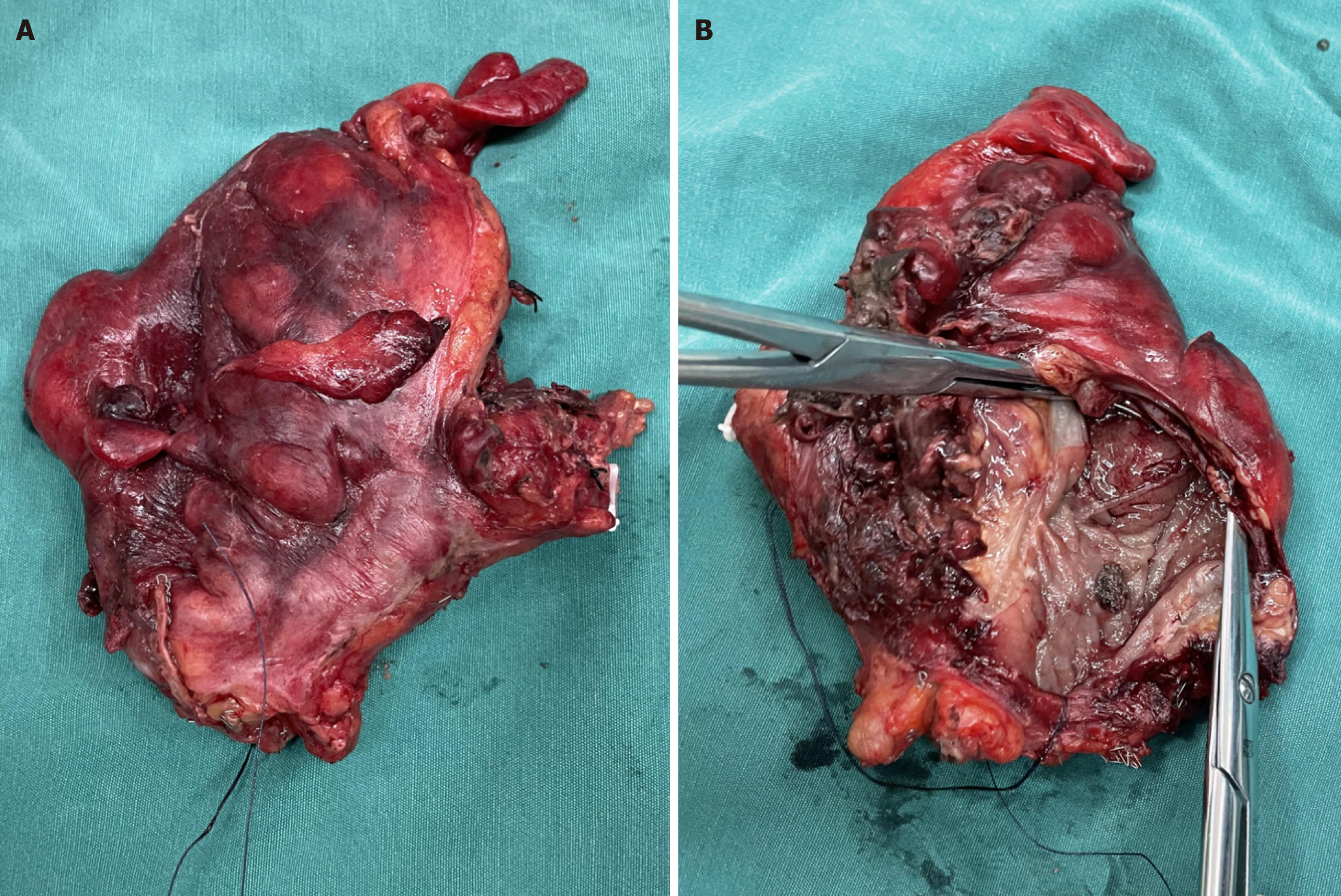
Figure 6 Photograph of the resected rectum.
A: Macroscopic appearance of the rectal surgery specimen; B: The appearance of the luminal cross-section of the incised rectal wall shows that on one side of the intestinal wall, there was visible congestion, erosion, and edematous tissue, with purulent necrotic secretions attached, and a perforation approximately 0.8 cm in size in the intestinal wall.
- Citation: Li F, Zhao B, Liu YQ, Chen GQ, Qu RF, Xu C, Long Z, Wu JS, Xiong M, Liu WH, Zhu L, Feng XL, Zhang L. Hematochezia due to rectal invasion by an internal iliac artery aneurysm: A case report. World J Clin Cases 2024; 12(11): 1980-1989
- URL: https://www.wjgnet.com/2307-8960/full/v12/i11/1980.htm
- DOI: https://dx.doi.org/10.12998/wjcc.v12.i11.1980


