Copyright
©The Author(s) 2024.
World J Clin Cases. Apr 16, 2024; 12(11): 1980-1989
Published online Apr 16, 2024. doi: 10.12998/wjcc.v12.i11.1980
Published online Apr 16, 2024. doi: 10.12998/wjcc.v12.i11.1980
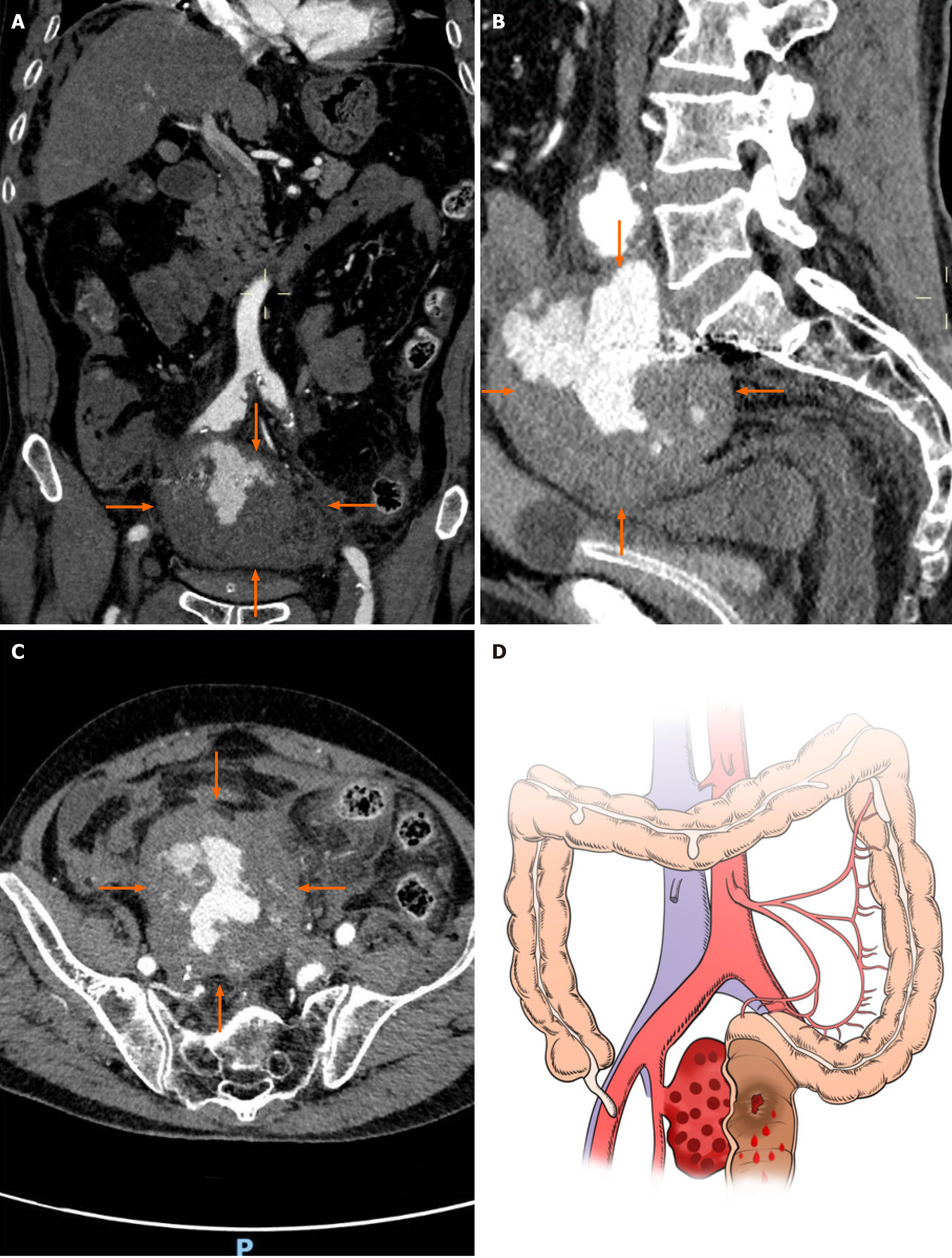
Figure 1 Computed tomography images of the abdomen and pelvis.
A: Coronal computed tomography (CT) image showing an oval-shaped mass with mixed high and low densities near the bifurcation of the right internal iliac artery (orange arrow); B: Coronal CT image showing an internal iliac aneurysm occupying the intrapelvic space, with a vertical diameter of approximately 7 centimeters, and suspected to be communicating with the rectum; C: Axial CT image showing that the internal iliac artery aneurysm had transverse and anteroposterior diameters of approximately 9.5 cm and 8.5 cm, respectively, suggesting the possibility of aneurysm rupture and hematoma formation; D: The illustration shows that the rectal wall was invaded by the right internal iliac artery aneurysm, which led to rectal bleeding.
- Citation: Li F, Zhao B, Liu YQ, Chen GQ, Qu RF, Xu C, Long Z, Wu JS, Xiong M, Liu WH, Zhu L, Feng XL, Zhang L. Hematochezia due to rectal invasion by an internal iliac artery aneurysm: A case report. World J Clin Cases 2024; 12(11): 1980-1989
- URL: https://www.wjgnet.com/2307-8960/full/v12/i11/1980.htm
- DOI: https://dx.doi.org/10.12998/wjcc.v12.i11.1980


