Copyright
©The Author(s) 2024.
World J Clin Cases. Apr 16, 2024; 12(11): 1960-1966
Published online Apr 16, 2024. doi: 10.12998/wjcc.v12.i11.1960
Published online Apr 16, 2024. doi: 10.12998/wjcc.v12.i11.1960
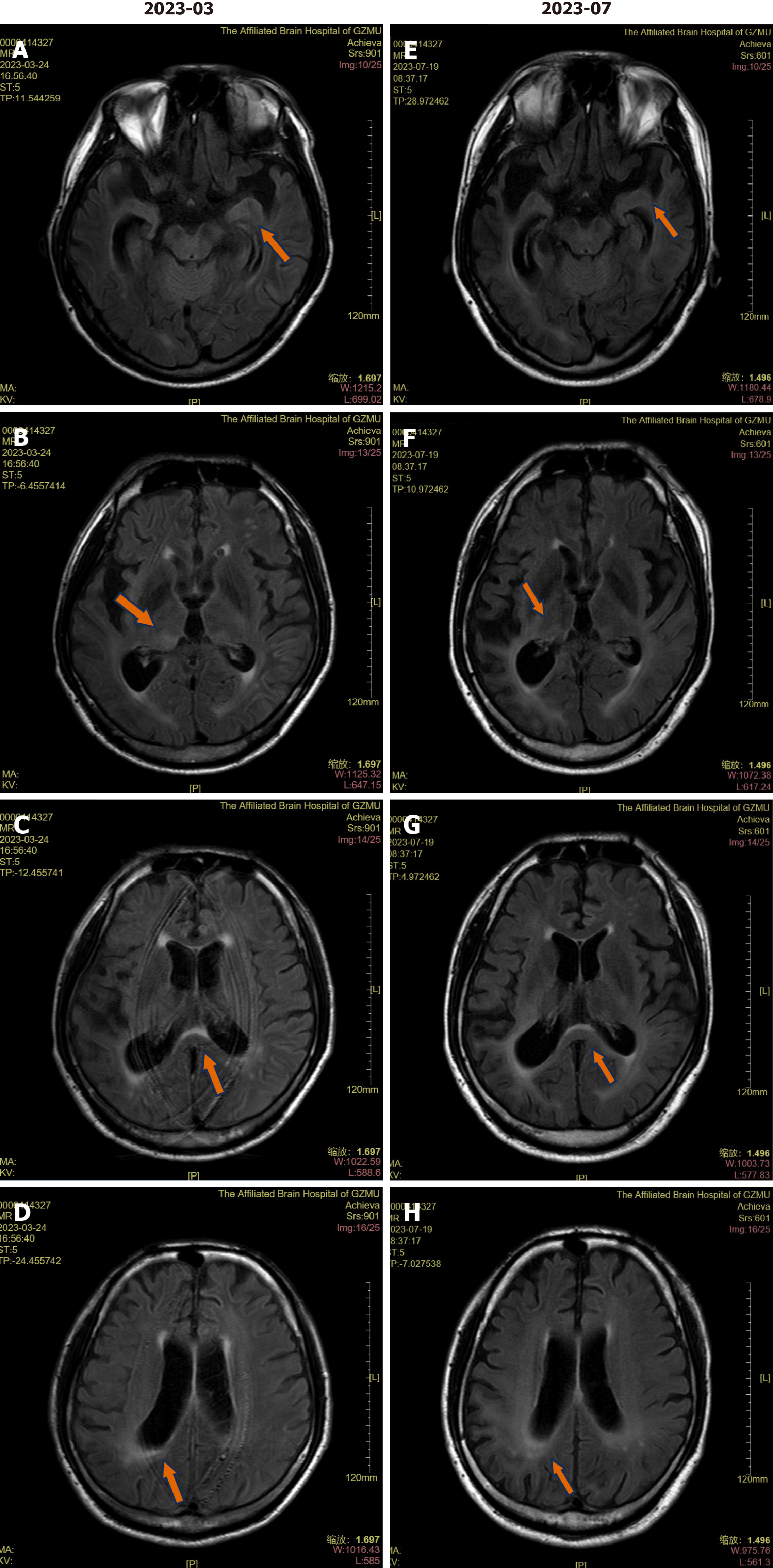
Figure 2 Brain magnetic resonance imaging scan on admission and after three months.
A and E: T2 fluid-attenuated inversion recovery (FLAIR) image shows the high signal of the left amygdala; B and F: T2 FLAIR image shows the high signal of the right thalamus; C and G: T2 FLAIR image shows the high signal of the corpus callosum; D and H: T2 FLAIR image shows the high signal in the white matter surrounding the posterior part of the lateral ventricular body.
- Citation: Fang YX, Zhou XM, Zheng D, Liu GH, Gao PB, Huang XZ, Chen ZC, Zhang H, Chen L, Hu YF. Neurosyphilis complicated by anti-γ-aminobutyric acid-B receptor encephalitis: A case report. World J Clin Cases 2024; 12(11): 1960-1966
- URL: https://www.wjgnet.com/2307-8960/full/v12/i11/1960.htm
- DOI: https://dx.doi.org/10.12998/wjcc.v12.i11.1960


