Copyright
©The Author(s) 2024.
World J Clin Cases. Apr 6, 2024; 12(10): 1824-1829
Published online Apr 6, 2024. doi: 10.12998/wjcc.v12.i10.1824
Published online Apr 6, 2024. doi: 10.12998/wjcc.v12.i10.1824
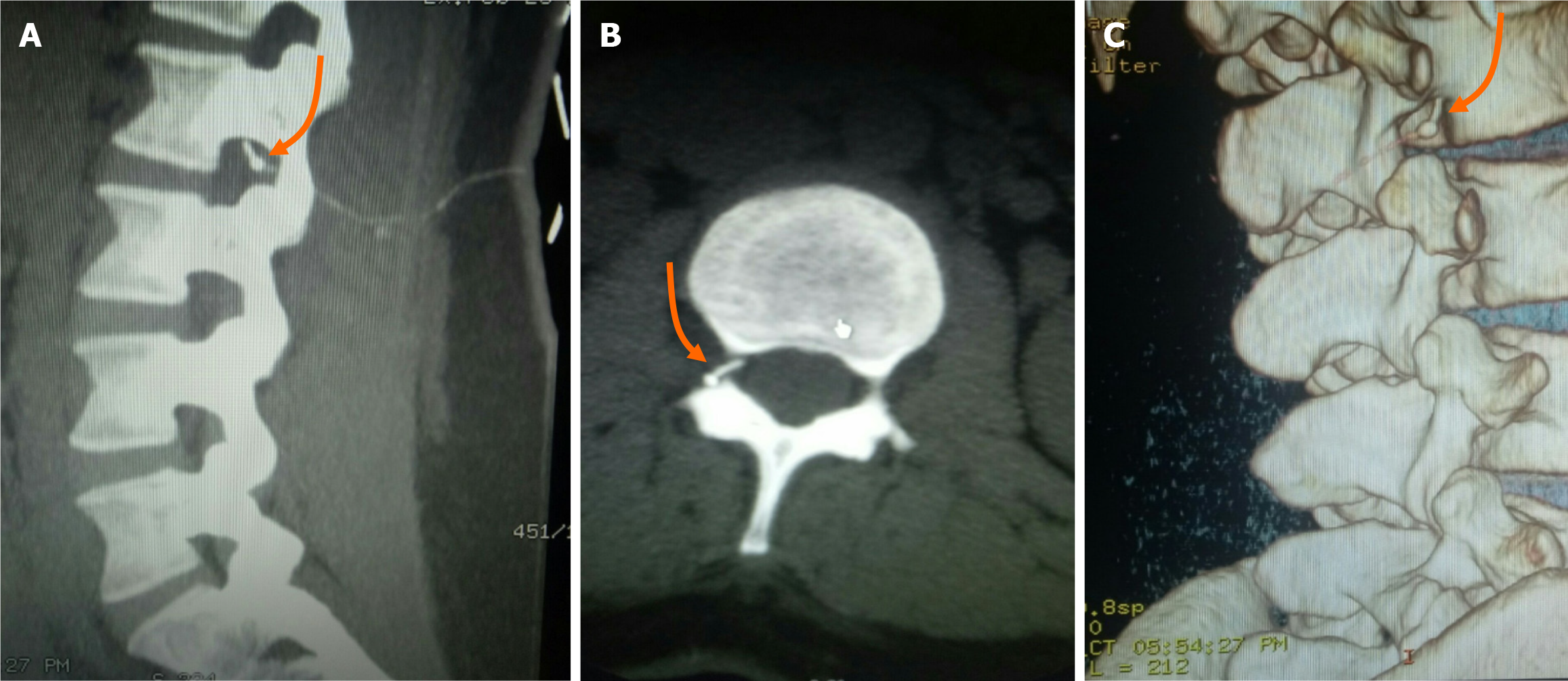
Figure 1 Computed tomography images of the patient.
A-C: Computed tomography images of the lumbar region show a knot in the catheter at the right subvertebral notch of the L2 vertebra (indicated by orange arrows).
- Citation: Deng NH, Chen XC, Quan SB. Unique method for removal of knotted lumbar epidural catheter: A case report. World J Clin Cases 2024; 12(10): 1824-1829
- URL: https://www.wjgnet.com/2307-8960/full/v12/i10/1824.htm
- DOI: https://dx.doi.org/10.12998/wjcc.v12.i10.1824


