Copyright
©The Author(s) 2024.
World J Clin Cases. Apr 6, 2024; 12(10): 1778-1784
Published online Apr 6, 2024. doi: 10.12998/wjcc.v12.i10.1778
Published online Apr 6, 2024. doi: 10.12998/wjcc.v12.i10.1778
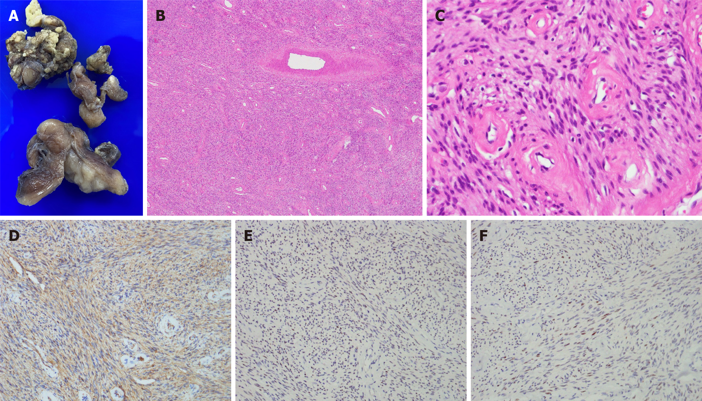
Figure 3 Pathological findings.
A: Grossly, the excised tumors were gray and elastic; B: Cellular angiofibroma displayed numerous thick-walled blood vessels with wall hyalinization (hematoxylin and eosin [H&E] 40 ×); C: The stroma contained small uniform short spindle-shaped cells with fusiform nuclei and pale indistinct cytoplasm (H&E 400 ×); D: Immunohistochemistry was positive for smooth muscle actin; E: Immunohistochemistry of the estrogen receptor showed focal positivity; F: Immunohistochemistry of the progesterone receptor showed focal positivity.
- Citation: Chen HE, Lu YY, Su RY, Wang HH, Chen CY, Hu JM, Kang JC, Lin KH, Pu TW. Cellular angiofibroma arising from the rectocutaneous fistula in an adult: A case report. World J Clin Cases 2024; 12(10): 1778-1784
- URL: https://www.wjgnet.com/2307-8960/full/v12/i10/1778.htm
- DOI: https://dx.doi.org/10.12998/wjcc.v12.i10.1778


