Copyright
©The Author(s) 2024.
World J Clin Cases. Jan 6, 2024; 12(1): 224-231
Published online Jan 6, 2024. doi: 10.12998/wjcc.v12.i1.224
Published online Jan 6, 2024. doi: 10.12998/wjcc.v12.i1.224
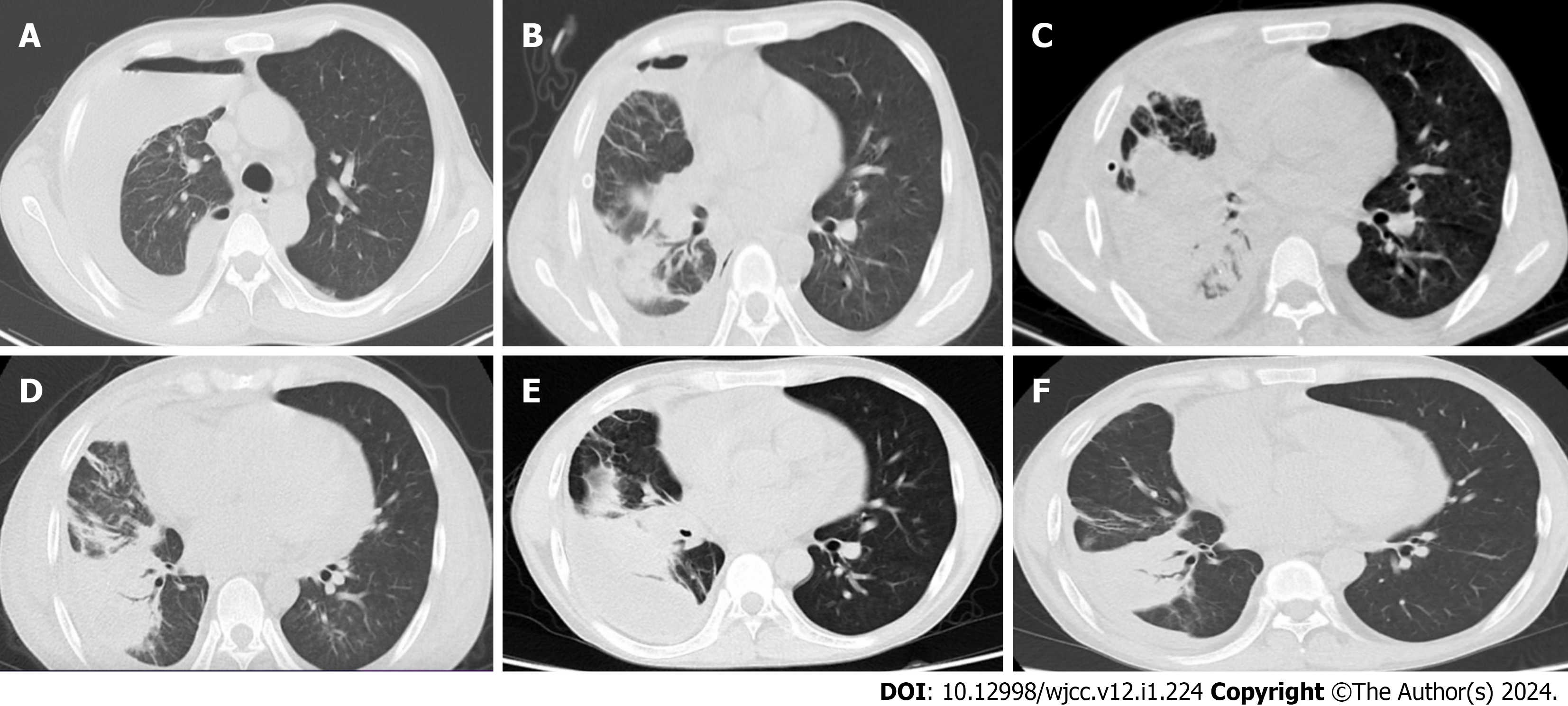
Figure 1 Chest computed tomography scan.
A: Right-sided fluid pneumothorax with multiple fluid within it; right lung with multiple exudates, right lower lobe abscesses; B: After drainage of right-sided fluid pneumothorax, the right-sided encapsulated effusion was significantly reduced compared with the previous one, and the pneumoperitoneum had been largely absorbed; the right intrapulmonary exudation and right lower lobe abscess were reduced compared with the previous one; the right middle lobe of the lung was atelectatic; C: Right pleural encapsulated effusion as before; right lower lobe abscess and multiple exudates in both lungs as before; right middle lobe atelectasis was slightly worse than before; D: Right pleural encapsulated effusion absorbed more than before; right lower lobe abscess and multiple right lung exudates reduced more than before; right middle lobe atelectasis as before; E: Right pleural encapsulated effusion was significantly greater than before; right lower lobe abscess and multiple right lung exudates were lesser than before; right middle lobe atelectasis was the same as before; F: Right pleural encapsulated effusion decreased compared to before; right lower lobe abscess and multiple right lung exudates as before; right middle lobe atelectasis as before.
- Citation: Liang GF, Chao S, Sun Z, Zhu KJ, Chen Q, Jia L, Niu YL. Pleural empyema with endobronchial mass due to Rhodococcus equi infection after renal transplantation: A case report and review of literature. World J Clin Cases 2024; 12(1): 224-231
- URL: https://www.wjgnet.com/2307-8960/full/v12/i1/224.htm
- DOI: https://dx.doi.org/10.12998/wjcc.v12.i1.224


