Copyright
©The Author(s) 2024.
World J Clin Cases. Jan 6, 2024; 12(1): 196-203
Published online Jan 6, 2024. doi: 10.12998/wjcc.v12.i1.196
Published online Jan 6, 2024. doi: 10.12998/wjcc.v12.i1.196
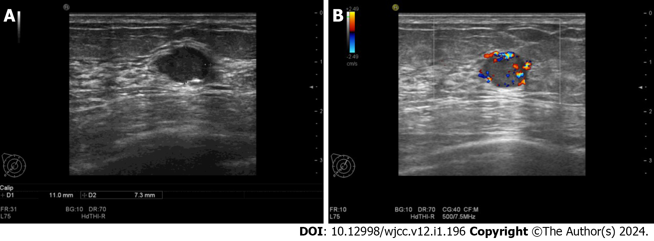
Figure 1 Ultrasound scan.
A: An 11 mm × 7 mm hypoechoic mass in the upper inner quadrant of the right breast, regular in shape, with a distinct margin; B: Color Doppler imaging showed hypervascularity.
- Citation: Ding JS, Zhang M, Zhou FF. Primary acinic cell carcinoma of the breast: A case report and review of literature. World J Clin Cases 2024; 12(1): 196-203
- URL: https://www.wjgnet.com/2307-8960/full/v12/i1/196.htm
- DOI: https://dx.doi.org/10.12998/wjcc.v12.i1.196


