Copyright
©The Author(s) 2024.
World J Clin Cases. Jan 6, 2024; 12(1): 169-175
Published online Jan 6, 2024. doi: 10.12998/wjcc.v12.i1.169
Published online Jan 6, 2024. doi: 10.12998/wjcc.v12.i1.169
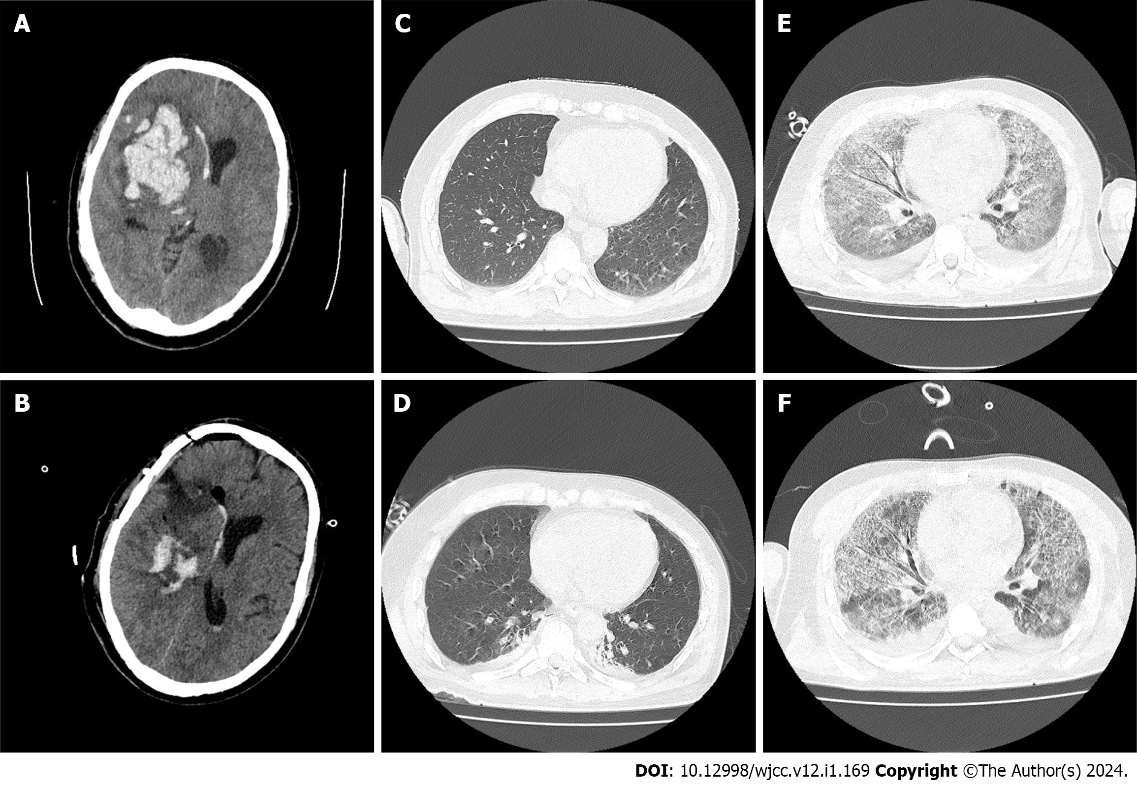
Figure 1 Computed tomography results.
A: Preoperative computed tomography (CT) findings. The hemorrhage was in the right basal ganglia, with a volume of approximately 60 mL. The brain tissue was pushed to the left, and the midline was obviously deviated to the left; B: Postoperative CT findings. The intracranial hematoma was basically cleared after surgery, and there was no obvious deviation in the midline, resulting in intracranial decompression; C and D: Before pulmonary infection with Elizabethkingia miricola, the lungs were in good condition at the time of admission to the emergency department and on the first day after surgery, without obvious inflammatory signs; E and F: Following pulmonary infection with Elizabethkingia miricola, lung CT showed diffuse distribution of a ground glass density shadow in both lungs, the air containing bronchial sign in local areas, thickening of bronchial vascular bundles in both lungs, and pulmonary edema.
- Citation: Qi PQ, Zeng YJ, Peng W, Kuai J. Lung imaging characteristics in a patient infected with Elizabethkingia miricola following cerebral hemorrhage surgery: A case report. World J Clin Cases 2024; 12(1): 169-175
- URL: https://www.wjgnet.com/2307-8960/full/v12/i1/169.htm
- DOI: https://dx.doi.org/10.12998/wjcc.v12.i1.169


