Copyright
©The Author(s) 2023.
World J Clin Cases. Mar 26, 2023; 11(9): 2091-2097
Published online Mar 26, 2023. doi: 10.12998/wjcc.v11.i9.2091
Published online Mar 26, 2023. doi: 10.12998/wjcc.v11.i9.2091
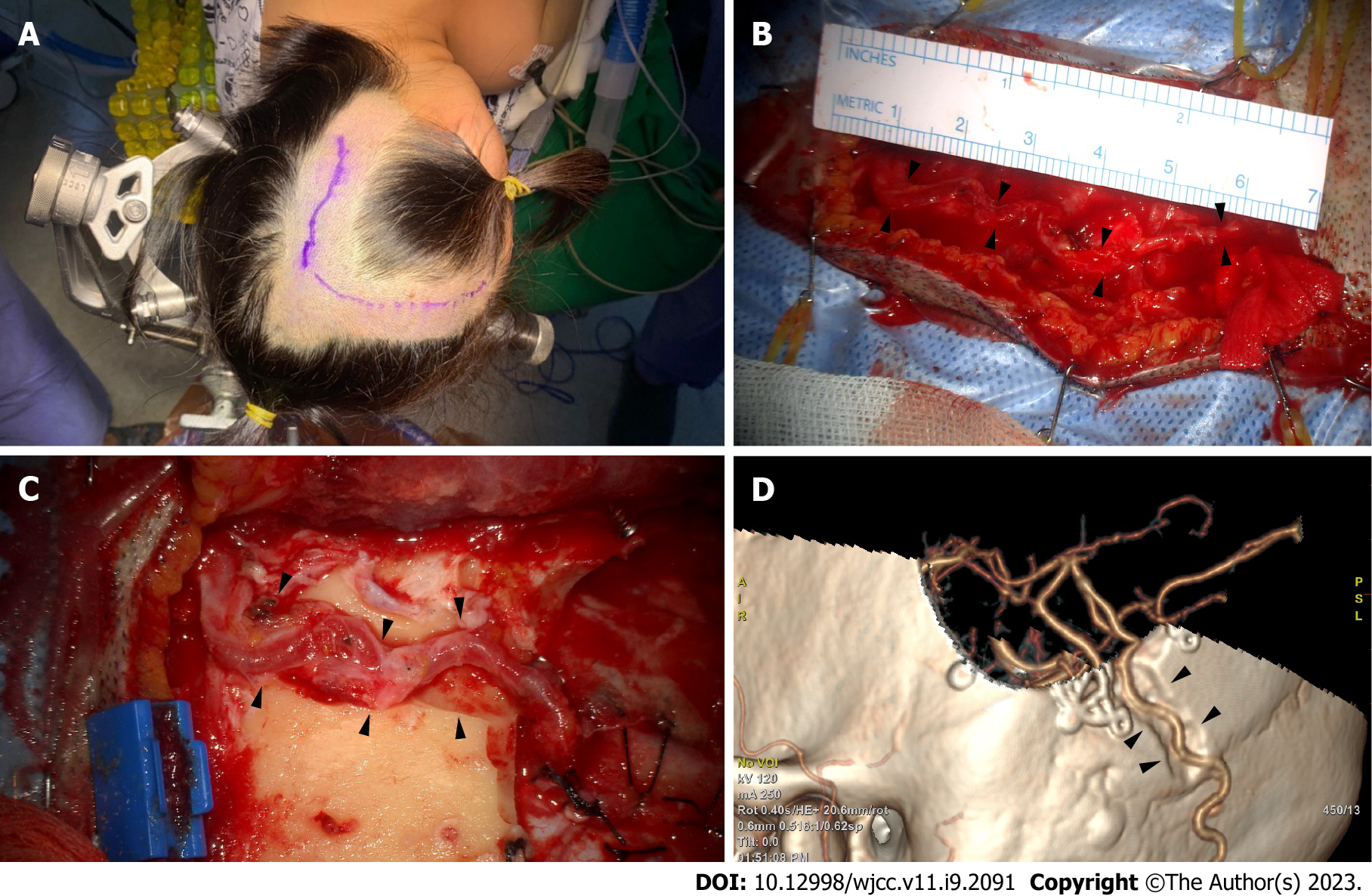
Figure 3 Occipital artery-middle cerebral artery bypass surgical position and incision.
A: In the three-quarter position, the retrosigmoid area was at the top, and the vertex was slightly raised. The occipital artery (OA) course was confirmed through angiography with fingertip palpating. An incision was created consecutively at the craniotomy site for the recipient artery; B: The OA’s main artery had a very tortuous course (black arrows); therefore, the soft tissue was dissected along the bend to secure the stretched curve’s full length to the recipient site, at least 7 cm; C and D: To prevent donor artery compression in the supine position, a route between the craniotomy site and OA’s proximal part was cut to create a sough (black arrows).
- Citation: Hong JH, Jung SC, Ryu HS, Kim TS, Joo SP. Occipital artery bypass importance in unsuitable superficial temporal artery: Two case reports. World J Clin Cases 2023; 11(9): 2091-2097
- URL: https://www.wjgnet.com/2307-8960/full/v11/i9/2091.htm
- DOI: https://dx.doi.org/10.12998/wjcc.v11.i9.2091


