Copyright
©The Author(s) 2023.
World J Clin Cases. Mar 26, 2023; 11(9): 2091-2097
Published online Mar 26, 2023. doi: 10.12998/wjcc.v11.i9.2091
Published online Mar 26, 2023. doi: 10.12998/wjcc.v11.i9.2091
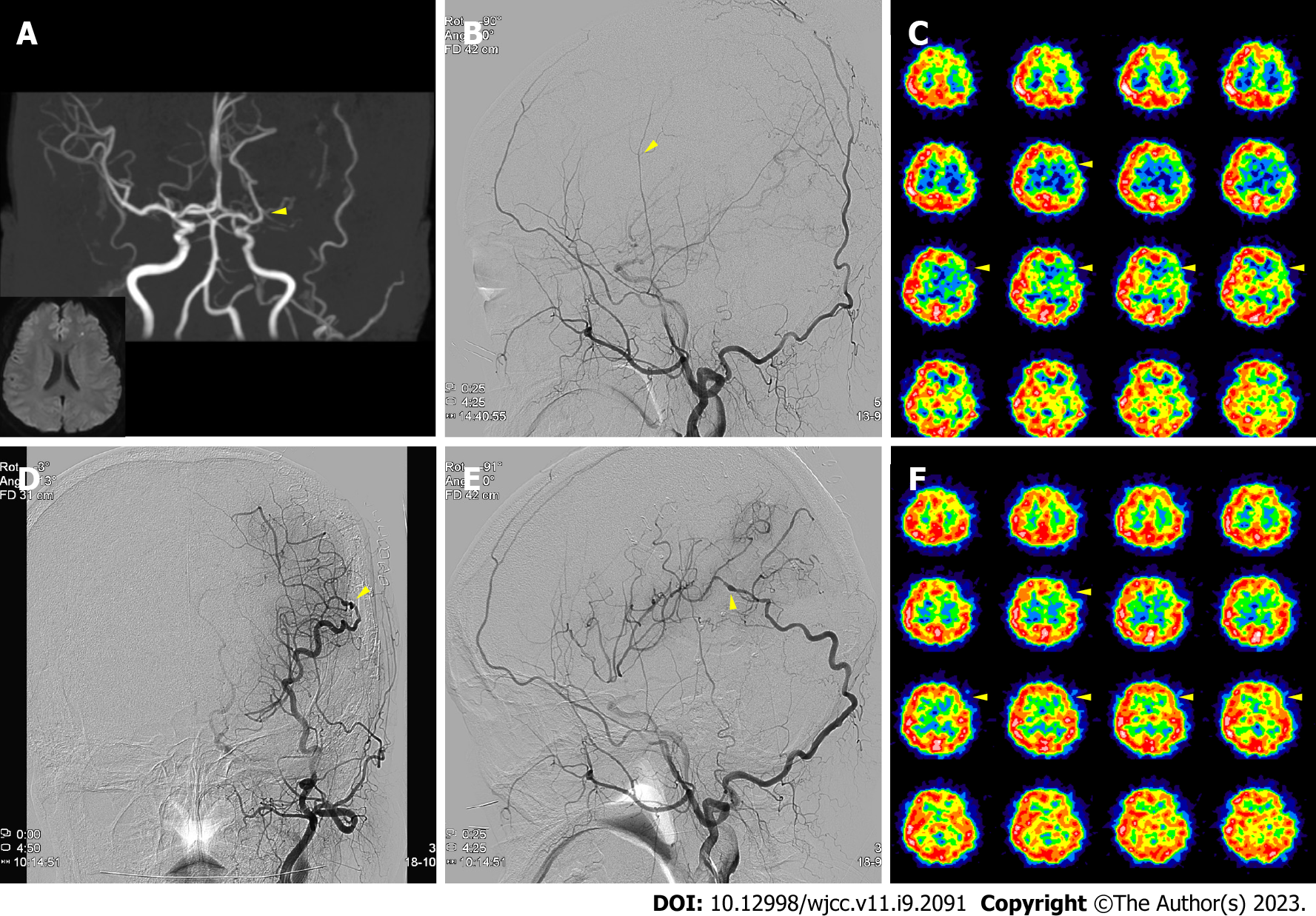
Figure 1 Case 1 perioperative imaging.
A: Initial magnetic resonance imaging indicated a small left frontal infarction from the left middle cerebral artery (MCA) stenosis (yellow arrow); B: On the transfemoral cerebral angiography (TFCA), the left superficial temporal artery was thin with weak flow (yellow arrow); however, the occipital artery (OA) was prominent (black arrows); C-F: After the OA-MCA bypass, blood flow at the surgical site was intact on postoperative TFCA (D and E; yellow arrow), with a perfusion defect improvement on postoperative Diamox SPECT compared with the preoperative scan (C and F; yellow arrows).
- Citation: Hong JH, Jung SC, Ryu HS, Kim TS, Joo SP. Occipital artery bypass importance in unsuitable superficial temporal artery: Two case reports. World J Clin Cases 2023; 11(9): 2091-2097
- URL: https://www.wjgnet.com/2307-8960/full/v11/i9/2091.htm
- DOI: https://dx.doi.org/10.12998/wjcc.v11.i9.2091


