Copyright
©The Author(s) 2023.
World J Clin Cases. Mar 26, 2023; 11(9): 2021-2028
Published online Mar 26, 2023. doi: 10.12998/wjcc.v11.i9.2021
Published online Mar 26, 2023. doi: 10.12998/wjcc.v11.i9.2021
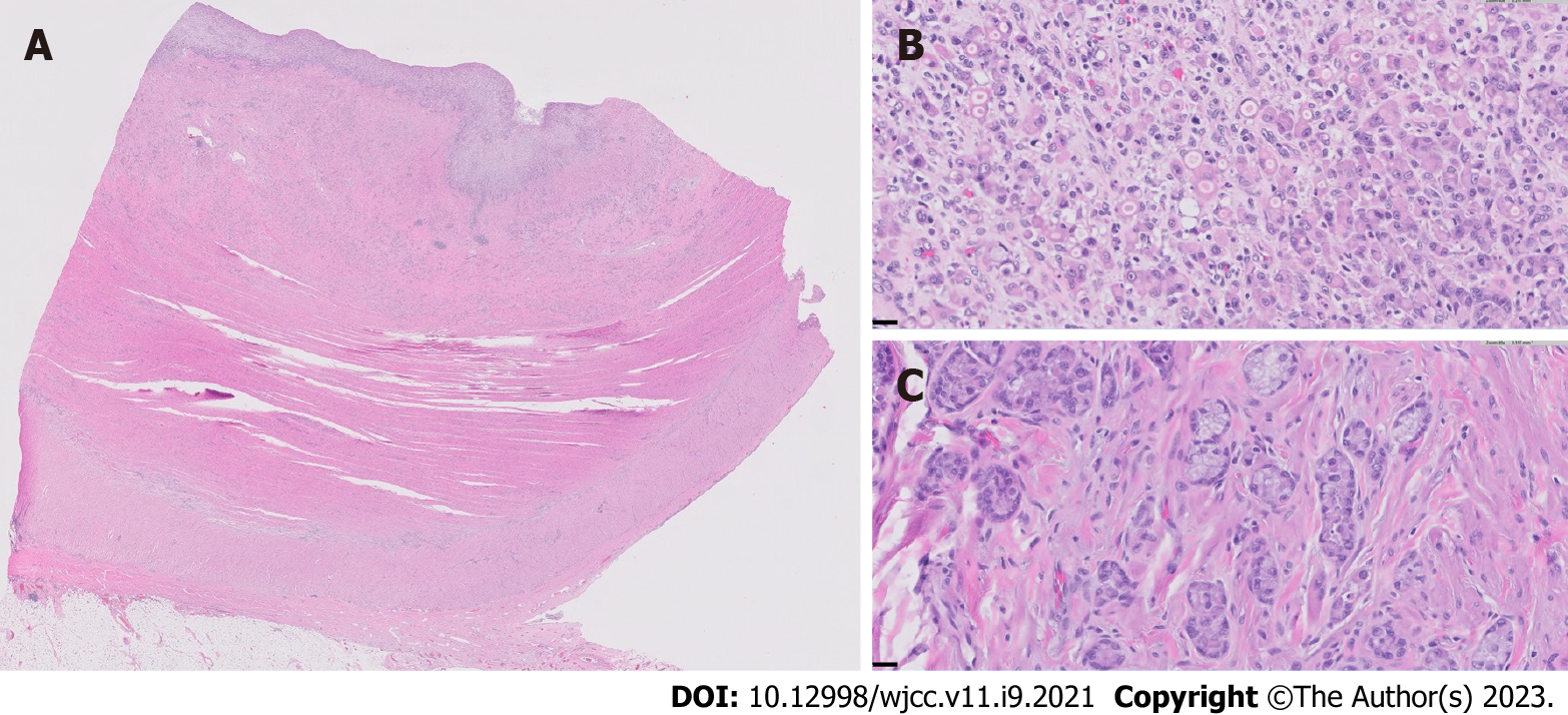
Figure 3 Small bowel adenocarcinoma in resected neoterminal ileum.
A: Cross section of the ileum near the anastomosis, showing complete effacement of the mucosa and infiltration of the underlying ileal wall by the adenocarcinoma (0.5 × objective magnification); B: Representative high-magnification view of the poorly-differentiated adenocarcinoma, comprised of sheets of single malignant cells and cell clusters with signet ring morphology (40 × objective magnification); C: Representative high-magnification view of a well-differentiated region of the adenocarcinoma, characterized by well-defined tubules with small, rigid lumina, mimicking intestinal crypts, as well as tumor cells arranged in well-polarized clusters without distinct lumina (40 × objective magnification).Tumor was noted in 6/35 lymph nodes and showed serosal involvement and lymphovascular and perineural invasion. Immunohistochemistry revealed intact expression of DNA mismatch proteins MLH1, MSH2, MSH6 and PMS2.
- Citation: Karthikeyan S, Shen J, Keyashian K, Gubatan J. Small bowel adenocarcinoma in neoterminal ileum in setting of stricturing Crohn’s disease: A case report and review of literature . World J Clin Cases 2023; 11(9): 2021-2028
- URL: https://www.wjgnet.com/2307-8960/full/v11/i9/2021.htm
- DOI: https://dx.doi.org/10.12998/wjcc.v11.i9.2021


