Copyright
©The Author(s) 2023.
World J Clin Cases. Mar 26, 2023; 11(9): 1992-2001
Published online Mar 26, 2023. doi: 10.12998/wjcc.v11.i9.1992
Published online Mar 26, 2023. doi: 10.12998/wjcc.v11.i9.1992
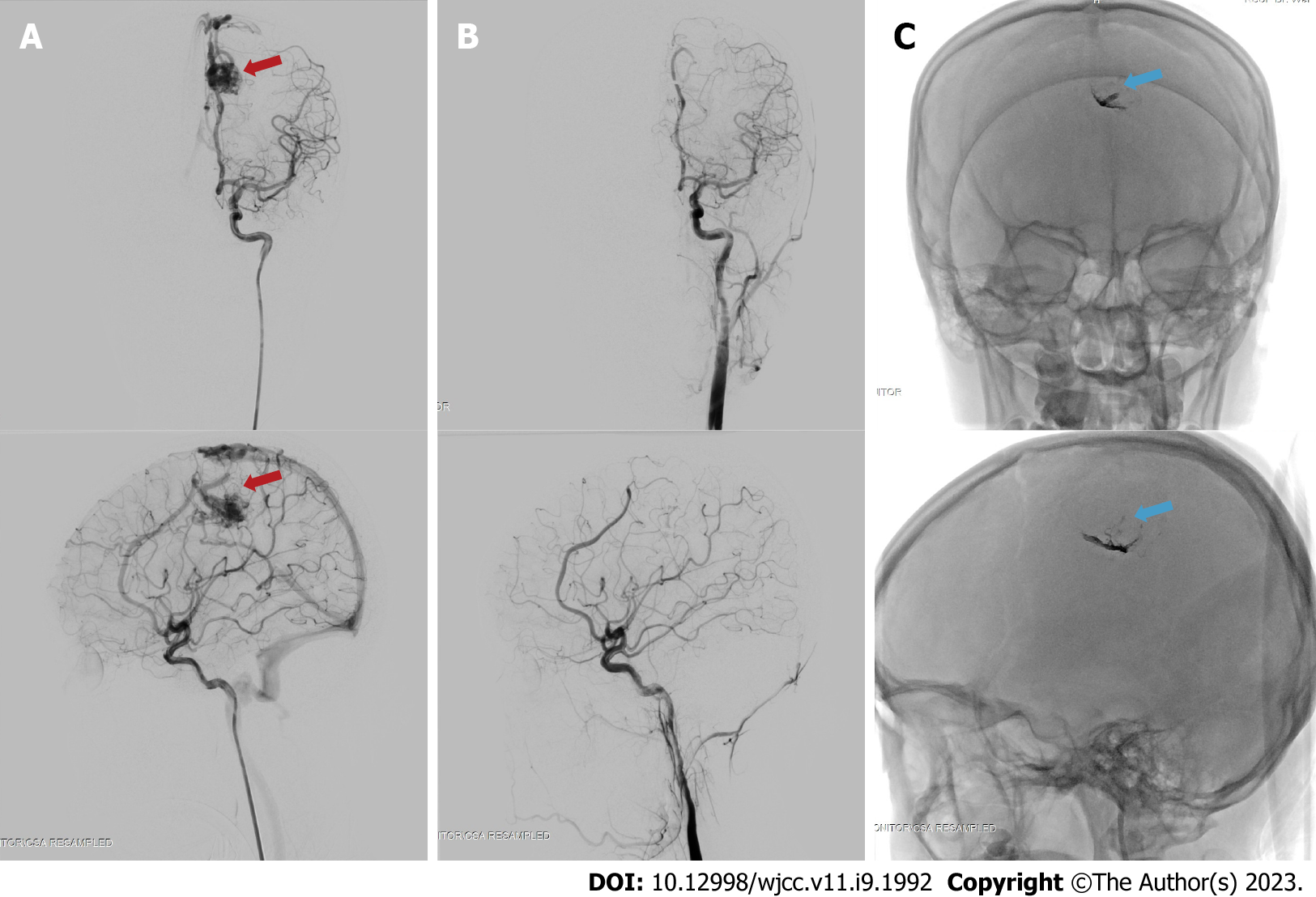
Figure 4 Digital subtraction angiography images before and after the embolization procedure on the brain arteriovenous malformation at approximately 3 mo post-ictus.
The embolization procedure was performed on the left pericallosal artery using the Onyx 18 until the nidus and draining vein can no longer be visualized. A: The nidus, before embolization (red arrow) on antero-posterior (top) and lateral (bottom) view of arterial phase; B: Complete obliteration of the nidus after embolization on the arterial phase, antero-posterior (top) and lateral (bottom) view; C: The onyx cast visible through fluoroscopy, on antero-posterior (top) and lateral (bottom) view, indicated with blue arrow.
- Citation: Bintang AK, Bahar A, Akbar M, Soraya GV, Gunawan A, Hammado N, Rachman ME, Ulhaq ZS. Delayed versus immediate intervention of ruptured brain arteriovenous malformations: A case report. World J Clin Cases 2023; 11(9): 1992-2001
- URL: https://www.wjgnet.com/2307-8960/full/v11/i9/1992.htm
- DOI: https://dx.doi.org/10.12998/wjcc.v11.i9.1992


