Copyright
©The Author(s) 2023.
World J Clin Cases. Mar 26, 2023; 11(9): 1992-2001
Published online Mar 26, 2023. doi: 10.12998/wjcc.v11.i9.1992
Published online Mar 26, 2023. doi: 10.12998/wjcc.v11.i9.1992
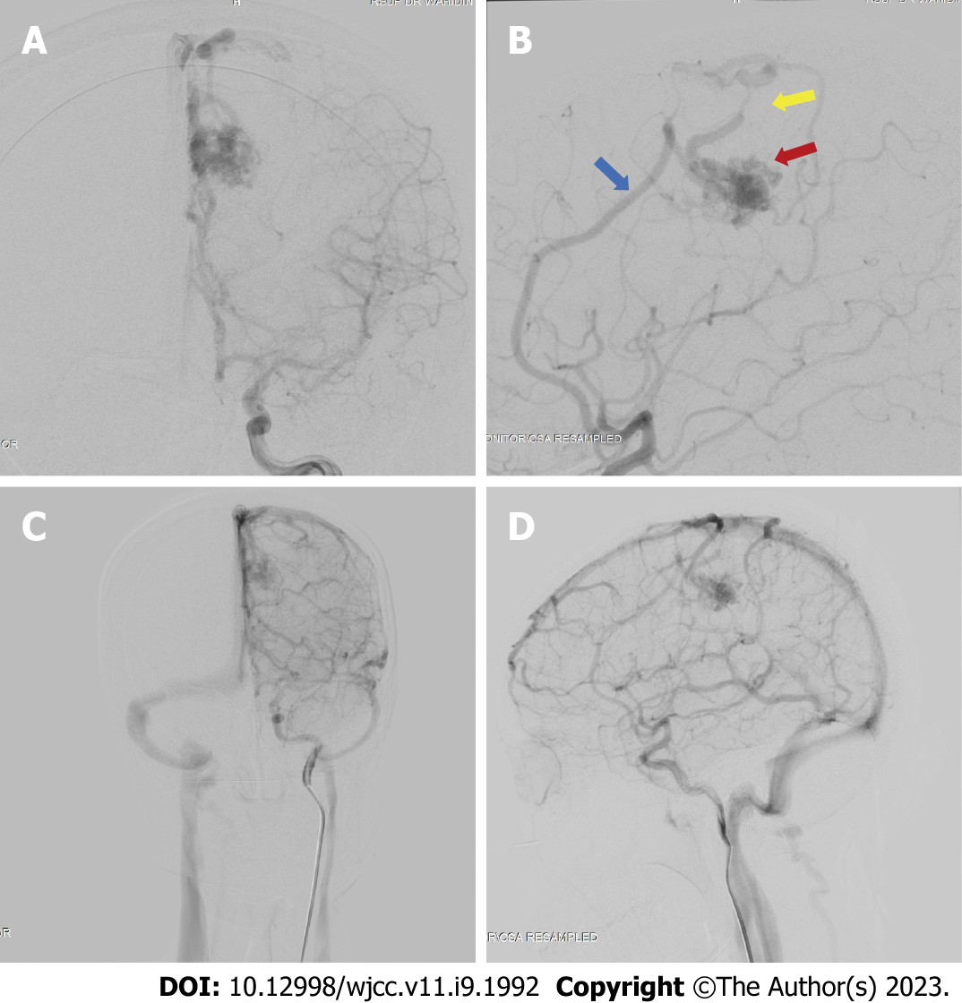
Figure 3 Digital subtraction angiography results of the patient.
The digital subtraction angiography image revealed the presence of an arteriovenous malformation (red arrow) feeding off the left pericallosal artery (blue arrow) and draining towards the cortical vein and a stenotic vein (yellow arrow). A: Late arterial phase anterior-posterior view; B: Late arterial phase, lateral view; C: Late venous phase, antero-posterior view; D: Late venous phase, lateral view.
- Citation: Bintang AK, Bahar A, Akbar M, Soraya GV, Gunawan A, Hammado N, Rachman ME, Ulhaq ZS. Delayed versus immediate intervention of ruptured brain arteriovenous malformations: A case report. World J Clin Cases 2023; 11(9): 1992-2001
- URL: https://www.wjgnet.com/2307-8960/full/v11/i9/1992.htm
- DOI: https://dx.doi.org/10.12998/wjcc.v11.i9.1992


