Published online Mar 26, 2023. doi: 10.12998/wjcc.v11.i9.2060
Peer-review started: December 2, 2022
First decision: February 2, 2023
Revised: February 10, 2023
Accepted: February 27, 2023
Article in press: February 27, 2023
Published online: March 26, 2023
Isolated capitate fractures are rare carpal fractures. Following high-energy injuries, capitate fractures are usually associated with other carpal fractures or ligament injuries. The management of capitate fractures depends on the fracture pattern. Here, we report an unusual capitate fracture with a dorsal shearing pattern and concomitant carpometacarpal dislocation, with a 6-year follow-up. To the best of our knowledge, this fracture pattern and surgical management have not been previously reported.
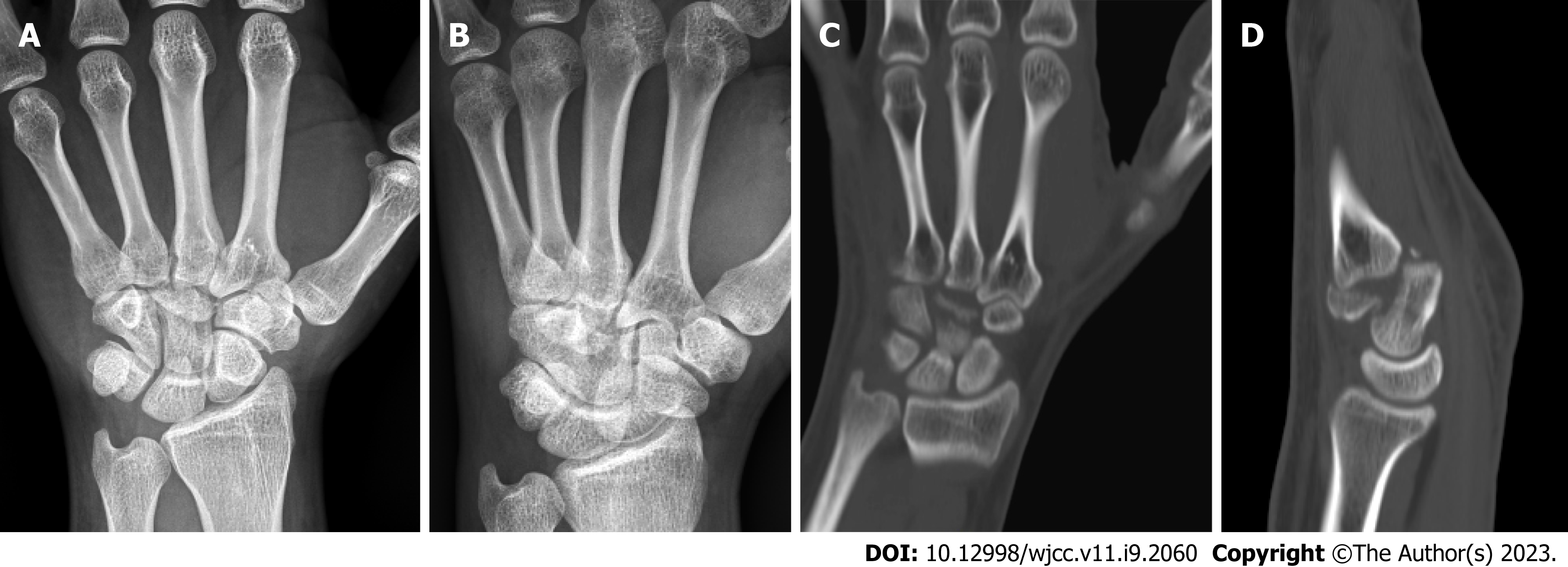
A 28-year-old man presented with left-hand volar tenderness and decreased grip strength that persisted for one month after a traffic accident. Radiography showed a distal capitate fracture with carpometacarpal joint incongruence. Computed tomography (CT) revealed a distal capitate fracture with carpometacarpal joint dislocation. The distal fragment was rotated by 90° in the sagittal plane, and an oblique shearing fracture pattern was noted. Open reduction and internal fixation (ORIF) with a locking plate were performed using the dorsal approach. The imaging studies performed 3 mo and 6 years following surgery revealed a healed fracture, and the Disabilities of the Arm, Shoulder, and Hand and visual analog scale scores were significantly improved.
CT can detect capitate fractures with dorsal shearing pattern and concomitant carpometacarpal dislocation. ORIF using a locking plate are possible.
Core Tip: Isolated capitate fractures with distal fragment rotated by 90° in the sagittal plane, dorsal shearing pattern and concomitant carpometacarpal dislocation have not been previously reported. Open reduction and internal fixation with a locking plate using the dorsal approach are possible. Long term radiological and clinical outcomes, including the Disabilities of the Arm, Shoulder, and Hand and visual analog scale scores were improved significantly.
- Citation: Lai CC, Fang HW, Chang CH, Pao JL, Chang CC, Chen YJ. Unusual capitate fracture with dorsal shearing pattern and concomitant carpometacarpal dislocation with a 6-year follow-up: A case report. World J Clin Cases 2023; 11(9): 2060-2066
- URL: https://www.wjgnet.com/2307-8960/full/v11/i9/2060.htm
- DOI: https://dx.doi.org/10.12998/wjcc.v11.i9.2060
Capitate fractures are rare and usually associated with other carpal bone fractures or ligament injuries. The reported incidence of these fractures is 0.42-4.9% of all adult carpal fractures[1-3]. The most frequent injury mechanism is a fall on an outstretched hand. With a greater energy injury mechanism, more neighboring structures may be injured. Greater arc perilunate fracture-dislocation is typically a high-energy injury with several bone fractures, including the scaphoid, capitate, and triquetrum. Scaphocapitate syndrome is a rare, greater arc fracture in which the proximal capitate fragment rotates 90°-180° in the sagittal plane[4,5]. The treatment of capitate fractures depends on the fracture pattern and associated injuries. Short-arm cast immobilization is indicated for isolated non-displaced capitate fractures. Displaced capitate fractures associated with injuries to neighboring structures, including carpal fractures and ligament tears, require surgical treatment[5,6]. Isolated capitate fractures have rarely been reported; when they have, fracture types have also been reported[5]. Most of these studies were small case series[3,5]. In this study, we report a case of capitate fracture with dorsal shearing fracture-dislocation of the distal pole that was treated using plate fixation. To our knowledge, this fracture pattern and surgical management have not been previously reported.
A 28-year-old man presented with left-hand pain that persisted for one month after he was hit by a car while riding a motorcycle.
Medical records were obtained and retrospectively examined after obtaining approval from the institutional review board of our hospital. Multiple injuries were found, including a brain concussion, spleen laceration, and left pelvic and femoral shaft fractures. The patient was taken to another hospital, where he received treatment. One month after discharge, he visited our clinic because of pain in his left hand.
There was no history of past illness.
There was no personal and family history.
During the physical examination, left carpal volar tenderness with decreased grip strength was noted.
Blood test results were within normal limit.
Radiography of the left hand showed a distal capitate fracture with carpometacarpal joint incongruence (Figure 1A and B). Computed tomography of the left hand revealed a distal capitate fracture with carpometacarpal joint dislocation (Figure 1C and D).
The patient was diagnosed with capitate fracture with dorsal shearing pattern and concomitant carpometacarpal dislocation of the left hand.
The patient underwent surgical treatment after serial examinations. Under general anesthesia, he was positioned supine with an arm table. Through a dorsal approach, a longitudinal skin incision was made over the 3rd carpometacarpal joint, followed by blunt dissection. The extensor tendon was identified and protected. The dorsal intercarpal ligament, distal fragment of the capitate fracture, and ruptured capsule were identified. After removing fibrotic tissue, the distal fragment of the capitate fracture was rotated by 90° in the sagittal plane, and an oblique shearing fracture pattern was observed. After reduction and repair of the capsule, we fixed the fracture using Strut Plate 1.3 (DePuy Synthes, Paoli, PA, United States), a mini-locking plate. The wound was closed in layer.
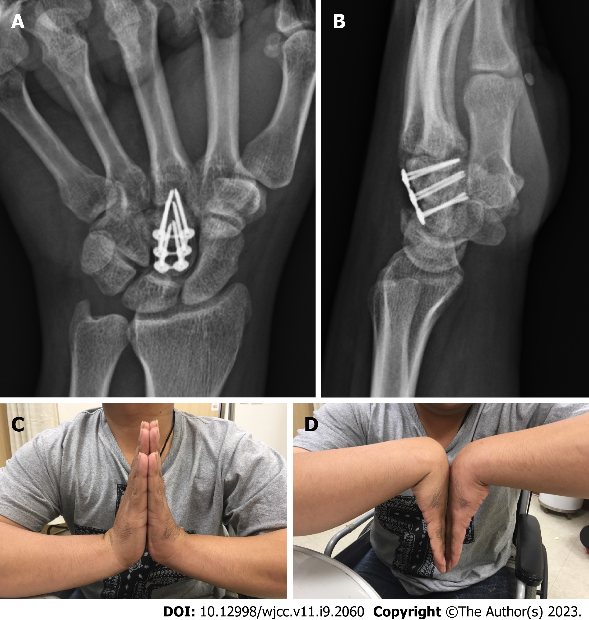
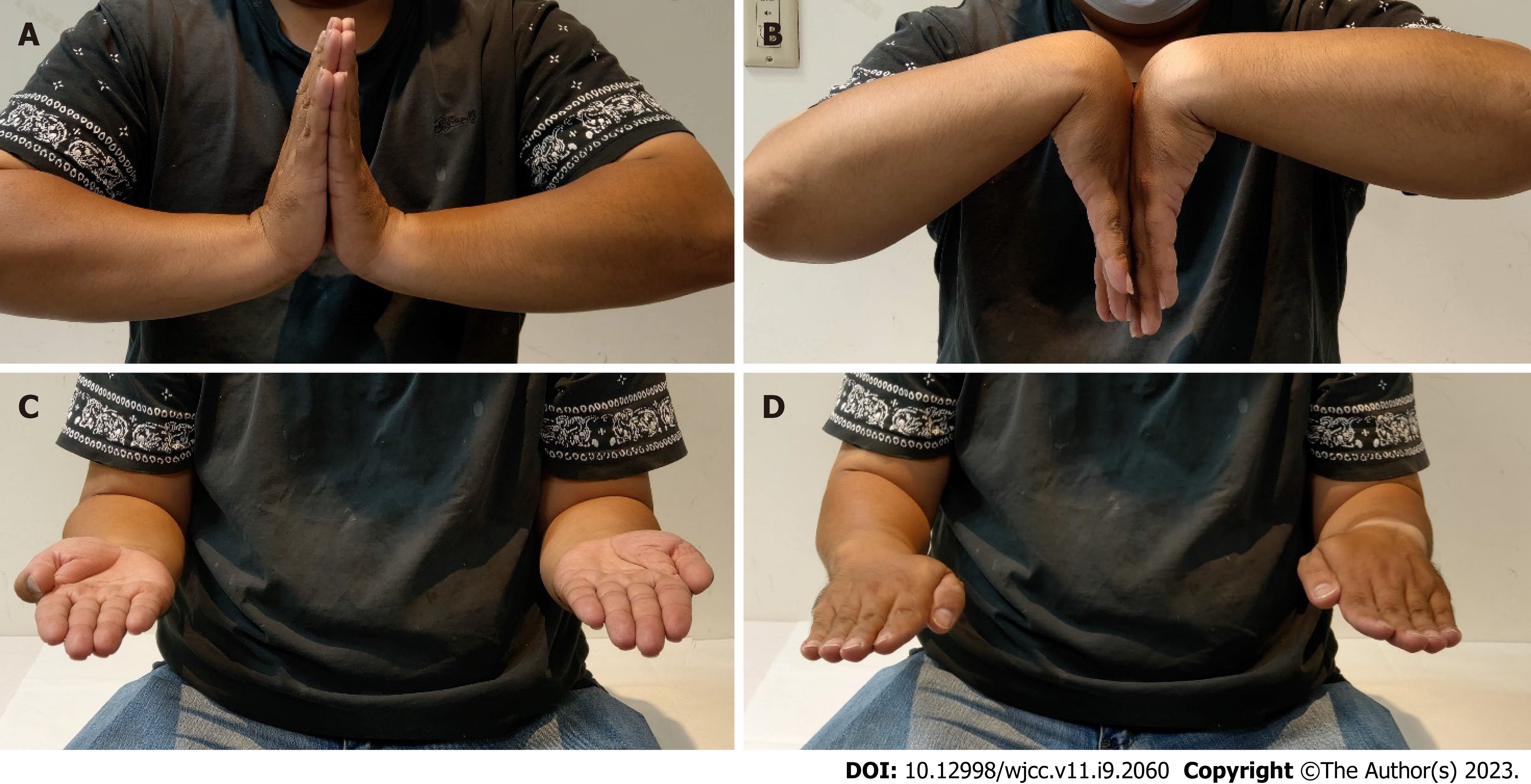
The patient was discharged three days after the operation. His hand was immobilized with a short-arm splint for 2 wk. Finger active range of motion rehabilitation were allowed immediately after operation. Wrist active range of motion and forearm rotation were allowed 2 wk postoperatively. Full weight-bearing was allowed 4 wk postoperatively, and he returned to his electronic technician job. The 3-month postoperative radiographs demonstrated appropriate plate and screw placement with no fracture site collapse (Figure 2A and B). His grip strength increased, and the left carpal volar tenderness resolved. The flexion/extension of the left wrist was comparable to that of the dominant right wrist (Figure 2C and D). Radiography and computed tomography performed 6 years later revealed a healed fracture with no avascular necrosis of the capitate (Figure 3). Figure 4 shows clinical photos of the left wrist six years postoperatively. The flexion/extension of the left wrist and forearm rotation are comparable to that of the dominant right wrist. Clinical outcomes were evaluated using the Disabilities of the Arm, Shoulder and Hand (DASH) and visual analog scale (VAS) scores at 3 mo and 6 years postoperatively. The DASH scores decreased from 45.0 to 18.3, while the VAS scores decreased from 3 to 0 at three months and six years postoperatively, respectively. No complications were noted in this patient.
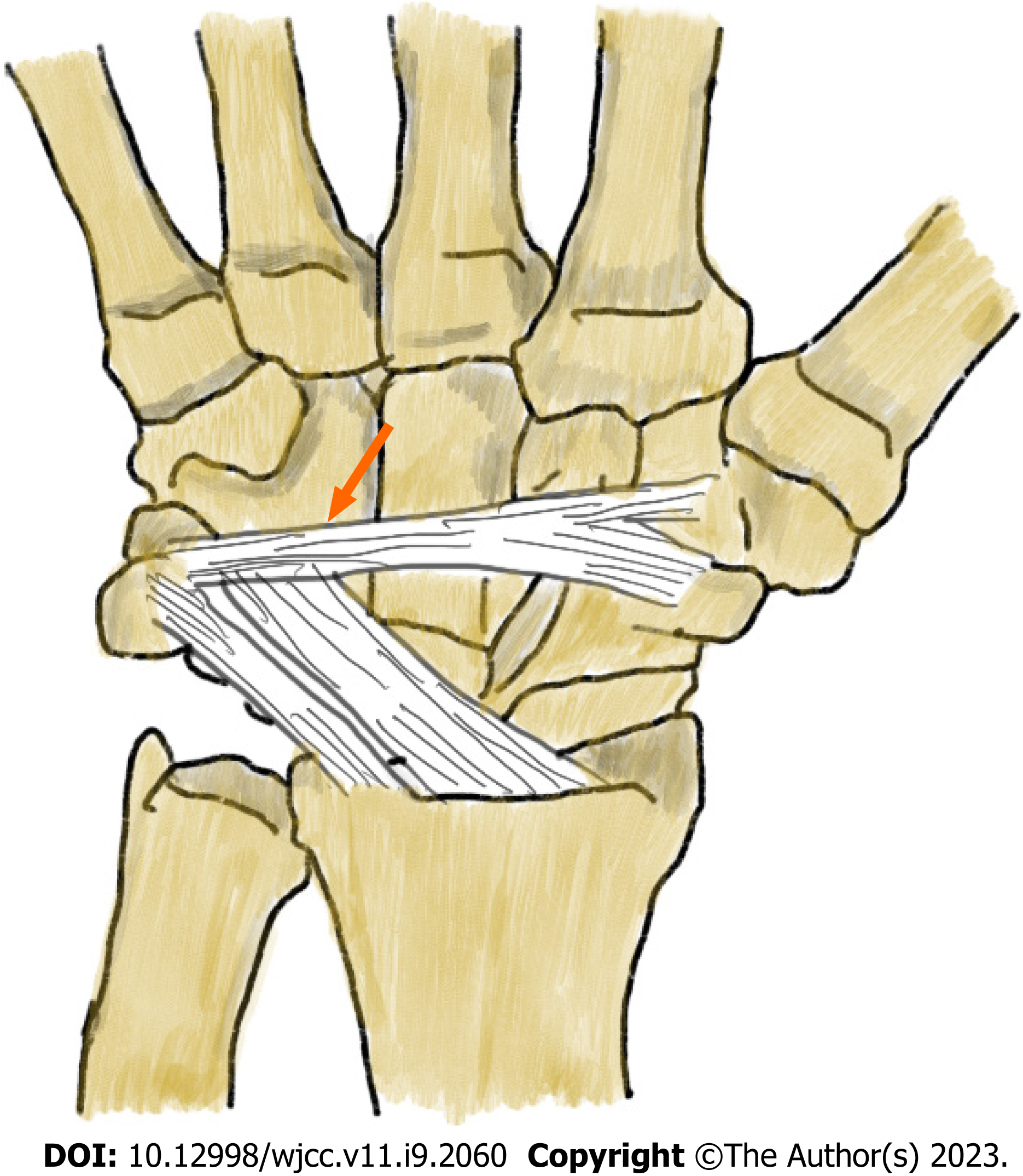
Four common capitate fracture patterns have been described: Transverse pole, transverse body, verticofrontal, and parasagittal[6]. Moran et al[5] reported the largest retrospective case series, which included 53 patients classified into three groups: capitate body (including stellate comminuted, oblique high and low, and transverse high and low), avulsion tip (including dorsal and volar tip avulsions), and shear depression (including coronal shear and dorsal depression) fractures. In our study, the patient presented with a dorsal shearing fracture of the distal pole with concomitant carpometacarpal dislocation and fracture fragment rotated by 90° in the sagittal plane. To our knowledge, this is the first such reported case. The common mechanism of injury is direct axial loading on the third metacarpal base with an outstretched hand. On the dorsal aspect, the dorsal intercarpal (DIC) and dorsal radiocarpal ligaments provide stability to the carpal bones, including the scaphoid and lunate[7]. In our study, the distal fragment of the capitate fracture was rotated by 90° in the sagittal plane with carpometacarpal dislocation. The possible mechanisms of injury leading to a 90° rotated fragment were blocking of the proximal part of the fragment by the DIC ligament and direct axial loading on the third metacarpal base with an extremely outstretched hand (Figure 5). Michael et al[8] reported the in vivo kinematics of the carpals during extreme wrist flexion and extension. In extreme extension, the capitate extends more than the scaphoid and lunate. The pressure between the dorsal aspect of the capitate and the DIC ligament also increases. As axial loading increases on the third metacarpal base, fracture-dislocation may occur.
The management of capitate fractures should be determined by the fracture location and concomitant injuries. The treatment options include surgical and non-surgical treatments. Open reduction and internal fixation with Kirschner wires or headless cannulated compression screws in displaced fractures and fracture dislocations have been reported[2,5,9,10]. For rigid fixation, stability and early rehabilitation, we used a locking plate to fix the unique fracture pattern, and the fracture was healed at the final follow-up. The most common complication after a capitate fracture is nonunion[3,5,6], with a reported nonunion rate of 19.6%[11]. Other complications have also been reported, including nonunion followed by avascular necrosis[12] and post-traumatic degenerative arthrosis of the mid-carpal joint[5]. In our case, no complications occurred. For diagnosis, physical examinations and radiographic images of the hand were important. If follow-up CT of the hand was available, the fracture pattern of capitate could have been precisely assessed and the surgical plan could be adequately prepared. To avoid nonunion complications, open reduction with rigid internal fixation with a locking plate could be an appropriate approach.
An unusual capitate fracture with a dorsal shearing fracture and concomitant carpometacarpal dislocation can be identified using computed tomography. Open reduction and internal fixation using locking plates are possible.
The authors thank the staff involved in the study at the Far Eastern Memorial Hospital, National Taipei University of Technology and Fu Jen Catholic University Hospital.
Provenance and peer review: Unsolicited article; Externally peer reviewed.
Peer-review model: Single blind
Specialty type: Orthopedics
Country/Territory of origin: Taiwan
Peer-review report’s scientific quality classification
Grade A (Excellent): 0
Grade B (Very good): 0
Grade C (Good): C, C
Grade D (Fair): 0
Grade E (Poor): 0
P-Reviewer: Cebula M, Poland; Yuan HJ, China S-Editor: Xing YX L-Editor: A P-Editor: Xing YX
| 1. | Adler JB, Shaftan GW. Fractures of the capitate. J Bone Joint Surg Am. 1962;44-A:1537-1547. [PubMed] [DOI] [Cited in This Article: ] |
| 2. | Freeland AE, Pesut TA. Oblique capitate fracture of the wrist. Orthopedics. 2004;27:287-290. [PubMed] [DOI] [Cited in This Article: ] [Cited by in Crossref: 5] [Cited by in F6Publishing: 7] [Article Influence: 0.4] [Reference Citation Analysis (0)] |
| 3. | Rand JA, Linscheid RL, Dobyns JH. Capitate fractures: a long-term follow-up. Clin Orthop Relat Res. 1982;209-216. [PubMed] [DOI] [Cited in This Article: ] |
| 4. | Monahan PR, Galasko CS. The scapho-capitate fracture syndrome. A mechanism of injury. J Bone Joint Surg Br. 1972;54:122-124. [PubMed] [Cited in This Article: ] |
| 5. | Kadar A, Morsy M, Sur YJ, Akdag O, Moran SL. Capitate Fractures: A Review of 53 Patients. J Hand Surg Am. 2016;41:e359-e366. [PubMed] [DOI] [Cited in This Article: ] [Cited by in Crossref: 18] [Cited by in F6Publishing: 16] [Article Influence: 2.0] [Reference Citation Analysis (0)] |
| 6. | Suh N, Ek ET, Wolfe SW. Carpal fractures. J Hand Surg Am. 2014;39:785-91; quiz 791. [PubMed] [DOI] [Cited in This Article: ] [Cited by in Crossref: 66] [Cited by in F6Publishing: 66] [Article Influence: 6.6] [Reference Citation Analysis (0)] |
| 7. | Mitsuyasu H, Patterson RM, Shah MA, Buford WL, Iwamoto Y, Viegas SF. The role of the dorsal intercarpal ligament in dynamic and static scapholunate instability. J Hand Surg Am. 2004;29:279-288. [PubMed] [DOI] [Cited in This Article: ] [Cited by in Crossref: 146] [Cited by in F6Publishing: 124] [Article Influence: 6.2] [Reference Citation Analysis (0)] |
| 8. | Rainbow MJ, Kamal RN, Leventhal E, Akelman E, Moore DC, Wolfe SW, Crisco JJ. In vivo kinematics of the scaphoid, lunate, capitate, and third metacarpal in extreme wrist flexion and extension. J Hand Surg Am. 2013;38:278-288. [PubMed] [DOI] [Cited in This Article: ] [Cited by in Crossref: 53] [Cited by in F6Publishing: 59] [Article Influence: 5.4] [Reference Citation Analysis (0)] |
| 9. | Sabat D, Arora S, Dhal A. Isolated capitate fracture with dorsal dislocation of proximal pole: a case report. Hand (N Y). 2011;6:333-336. [PubMed] [DOI] [Cited in This Article: ] [Cited by in Crossref: 15] [Cited by in F6Publishing: 14] [Article Influence: 1.1] [Reference Citation Analysis (0)] |
| 10. | Yoshihara M, Sakai A, Toba N, Okimoto N, Shimokobe T, Nakamura T. Nonunion of the isolated capitate waist fracture. J Orthop Sci. 2002;7:578-580. [PubMed] [DOI] [Cited in This Article: ] [Cited by in Crossref: 28] [Cited by in F6Publishing: 29] [Article Influence: 1.4] [Reference Citation Analysis (0)] |
| 11. | Rico AA, Holguin PH, Martin JG. Pseudarthrosis of the capitate. J Hand Surg Br. 1999;24:382-384. [PubMed] [DOI] [Cited in This Article: ] [Cited by in Crossref: 12] [Cited by in F6Publishing: 12] [Article Influence: 0.5] [Reference Citation Analysis (0)] |
| 12. | Vander Grend R, Dell PC, Glowczewskie F, Leslie B, Ruby LK. Intraosseous blood supply of the capitate and its correlation with aseptic necrosis. J Hand Surg Am. 1984;9:677-683. [PubMed] [DOI] [Cited in This Article: ] [Cited by in Crossref: 70] [Cited by in F6Publishing: 68] [Article Influence: 1.7] [Reference Citation Analysis (0)] |


