Copyright
©The Author(s) 2023.
World J Clin Cases. Mar 16, 2023; 11(8): 1878-1887
Published online Mar 16, 2023. doi: 10.12998/wjcc.v11.i8.1878
Published online Mar 16, 2023. doi: 10.12998/wjcc.v11.i8.1878
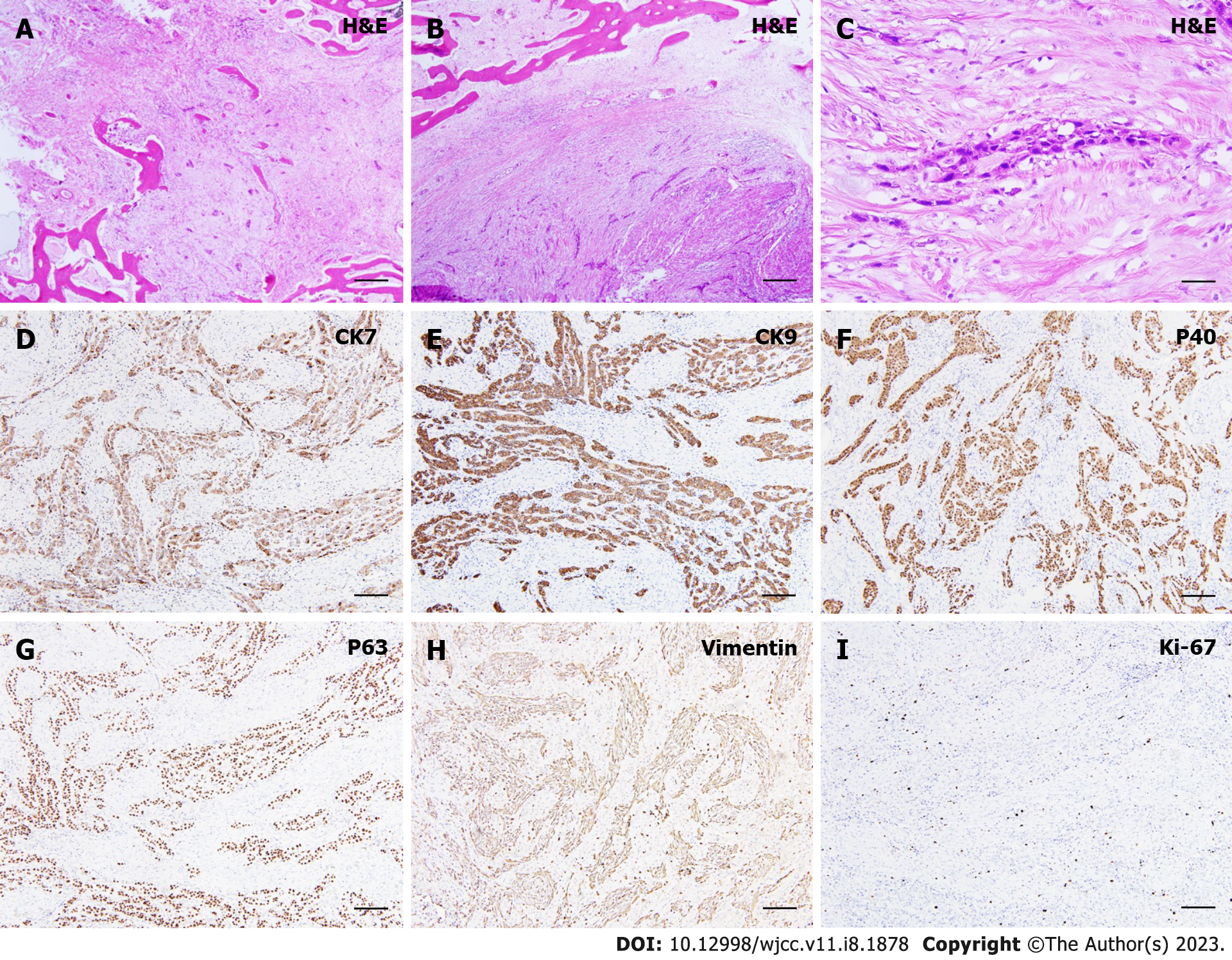
Figure 2 Histological findings of the present case.
A and B: Histological view focusing on small cords of epithelial cells of odontogenic origin immersed in a diffused sclerotic and collagenous stroma [hematoxylin and eosin (H&E), original magnification: × 20]; C: Evidence of vascular invasion (H&E, original magnification: × 20); D: Focal positivity in tumor cells to immunohistochemical staining for cytokeratin 7 (CK7) (original magnification: × 20); E: Immunohistochemistry for expression of CK19 demonstrating diffuse, uniform positivity in the neoplastic cells (original magnification: × 20); F: Immunohistochemistry demonstrating that the tumor cells expressed p40 (original magnification: × 20); G: Strong, diffuse positivity was seen for the expression of p63 (original magnification: × 20); H: The tumor cells were stained negative for vimentin (original magnification: × 20); I: Low proliferative activity (Ki-67) was seen (approximately 5%-10%) (original magnification: × 20). CK: Cytokeratin; H&E: Hematoxylin and eosin.
- Citation: Soh HY, Zhang WB, Yu Y, Zhang R, Chen Y, Gao Y, Peng X. Sclerosing odontogenic carcinoma of maxilla: A case report. World J Clin Cases 2023; 11(8): 1878-1887
- URL: https://www.wjgnet.com/2307-8960/full/v11/i8/1878.htm
- DOI: https://dx.doi.org/10.12998/wjcc.v11.i8.1878


