Copyright
©The Author(s) 2023.
World J Clin Cases. Mar 16, 2023; 11(8): 1878-1887
Published online Mar 16, 2023. doi: 10.12998/wjcc.v11.i8.1878
Published online Mar 16, 2023. doi: 10.12998/wjcc.v11.i8.1878
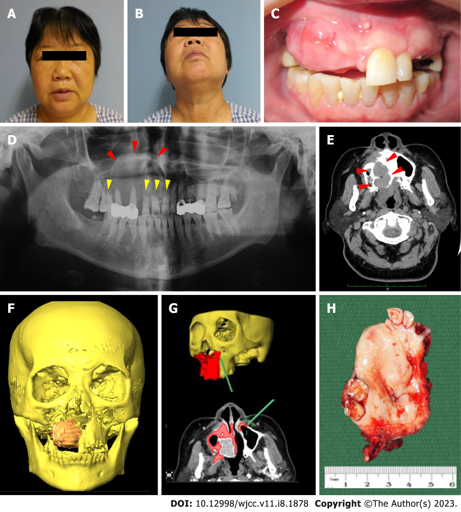
Figure 1 Clinical and radiological findings of the current case.
A and B: A thorough extra-oral and intra-oral examination was performed. The patient presented with a right facial swelling without overlying skin changes. The swelling was diffuse, firm, and non-tender, causing obliteration of the right nasolabial fold. Mouth opening was not restricted and there was no palpable cervical lymphadenopathy. There were no neurosensory changes to the right infraorbital region; C: Intra-oral examination showed an irregular mass at the anterior maxilla extending from the tooth 11 to 15 region and palatally crossing the midline into the left palate. There was no obliteration of the buccal sulcus. The swelling was firm and nontender, with a non-ulcerated overlying mucosa. The adjacent teeth showed no marked increase in mobility and no fluid discharge was noted on palpation of the swelling; D: The initial dental panoramic tomogram showed a radiolucent lesion of the right maxilla extending into the right maxillary sinus (red arrows) with marked root resorption (yellow arrows); E: Computed tomography scan with contrast showed an infiltrating lesion on the right maxilla with obvious bony destruction involving the right hard palate and right inferior turbinate (red arrows); F: Tumor mapping showing an expansile mass perforating both the buccal and palatal bone; G: The osteotomy lines were planned and confirmed using the intraoperative navigation system; H: Gross specimen of the lesion.
- Citation: Soh HY, Zhang WB, Yu Y, Zhang R, Chen Y, Gao Y, Peng X. Sclerosing odontogenic carcinoma of maxilla: A case report. World J Clin Cases 2023; 11(8): 1878-1887
- URL: https://www.wjgnet.com/2307-8960/full/v11/i8/1878.htm
- DOI: https://dx.doi.org/10.12998/wjcc.v11.i8.1878


