Copyright
©The Author(s) 2023.
World J Clin Cases. Mar 16, 2023; 11(8): 1808-1813
Published online Mar 16, 2023. doi: 10.12998/wjcc.v11.i8.1808
Published online Mar 16, 2023. doi: 10.12998/wjcc.v11.i8.1808
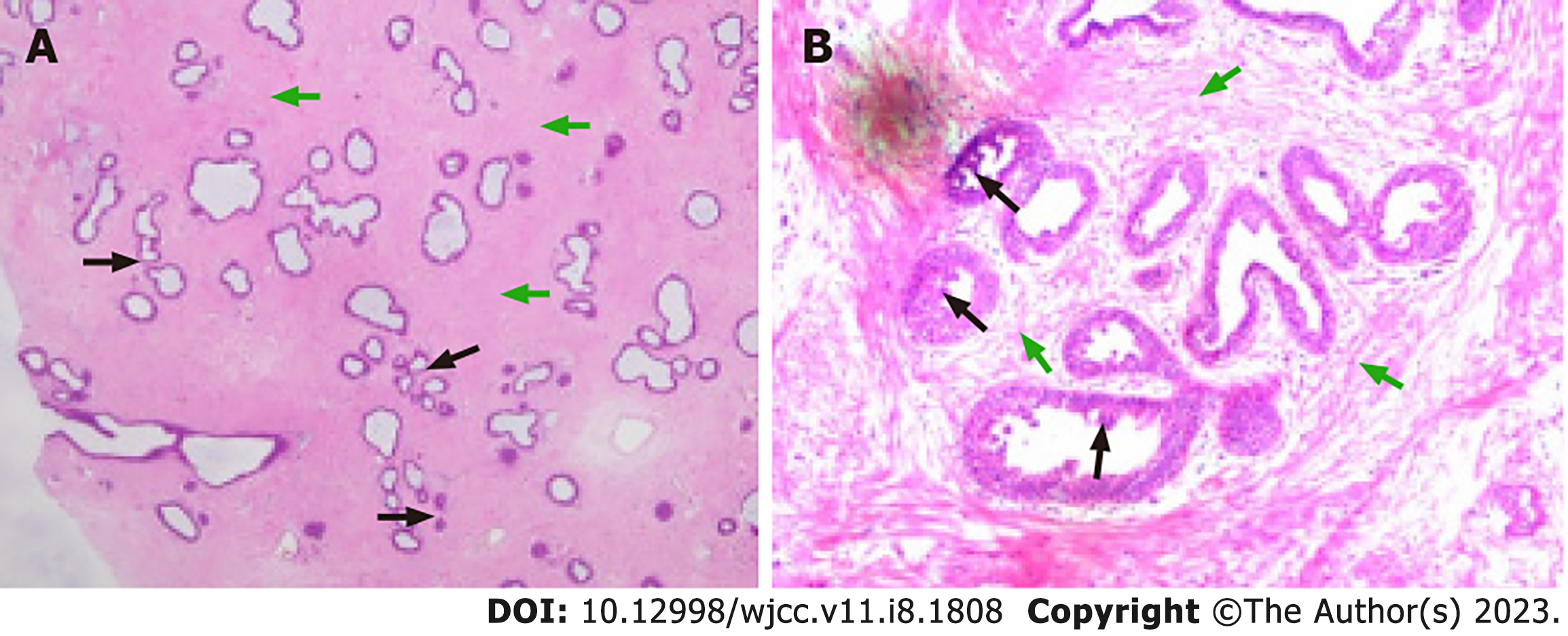
Figure 2 Pathological examination.
A: Low magnification (10 × 10 times) shows a massive ductal epithelial hyperplasia (black arrow) surrounded by a comparatively homogeneous stromal component (green arrow). The interstitial cells in the local lesions are dense, and there is no mitotic phase or cell abnormality; B: Low magnification (10 × 20 times) shows the columnar cells with punctate protrusion in the mammary ducts (black arrow). The density of stromal cells increased without obvious heteromorphism, which was mainly manifested by the interlaced cluster arrangement of fibroblasts and myofibroblasts (green arrow), showing the peritubular type.
- Citation: Wang J, Zhang DD, Cheng JM, Chen HY, Yang RJ. Giant juvenile fibroadenoma in a 14-year old Chinese female: A case report. World J Clin Cases 2023; 11(8): 1808-1813
- URL: https://www.wjgnet.com/2307-8960/full/v11/i8/1808.htm
- DOI: https://dx.doi.org/10.12998/wjcc.v11.i8.1808


