Copyright
©The Author(s) 2023.
World J Clin Cases. Mar 16, 2023; 11(8): 1782-1787
Published online Mar 16, 2023. doi: 10.12998/wjcc.v11.i8.1782
Published online Mar 16, 2023. doi: 10.12998/wjcc.v11.i8.1782
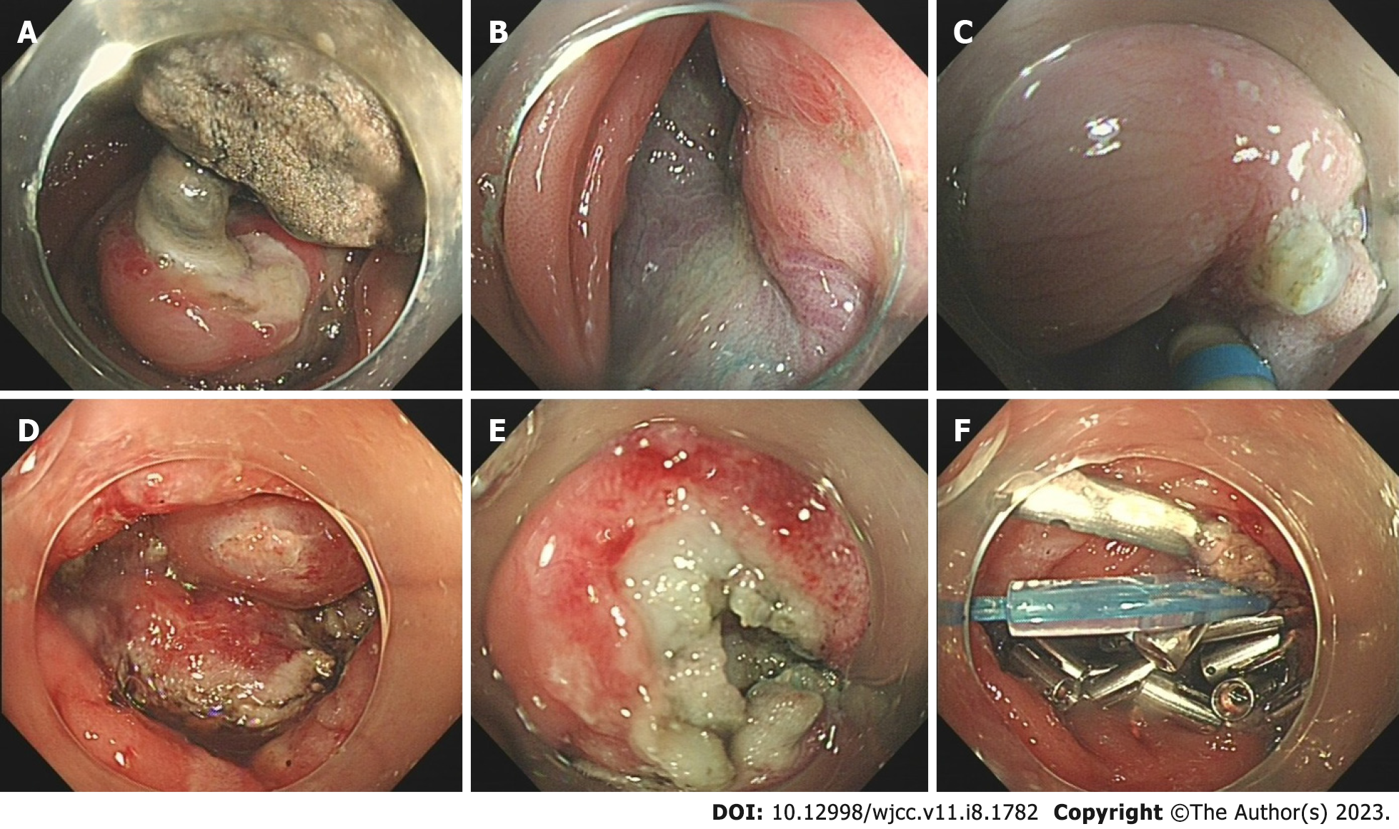
Figure 2 Endoscopy images.
A: The pedicle attached to the colon; B: Localized polyp patterns can be seen endoscopically; C: Circumferential pre-cutting around the lesion after submucosal injection; D: Endoscopic full-thickness resection of the polyp; E: Endoscopic transcanal incision; F: Closure of the wound using the purse-string closure method with nylon cord and multiple metallic clips.
- Citation: Ye L, Zhong JH, Liu YP, Chen DD, Ni SY, Peng FQ, Zhang S. Extensively infarcted giant solitary hamartomatous polyp treated with endoscopic full-thickness resection: A case report. World J Clin Cases 2023; 11(8): 1782-1787
- URL: https://www.wjgnet.com/2307-8960/full/v11/i8/1782.htm
- DOI: https://dx.doi.org/10.12998/wjcc.v11.i8.1782


