Copyright
©The Author(s) 2023.
World J Clin Cases. Mar 16, 2023; 11(8): 1771-1781
Published online Mar 16, 2023. doi: 10.12998/wjcc.v11.i8.1771
Published online Mar 16, 2023. doi: 10.12998/wjcc.v11.i8.1771
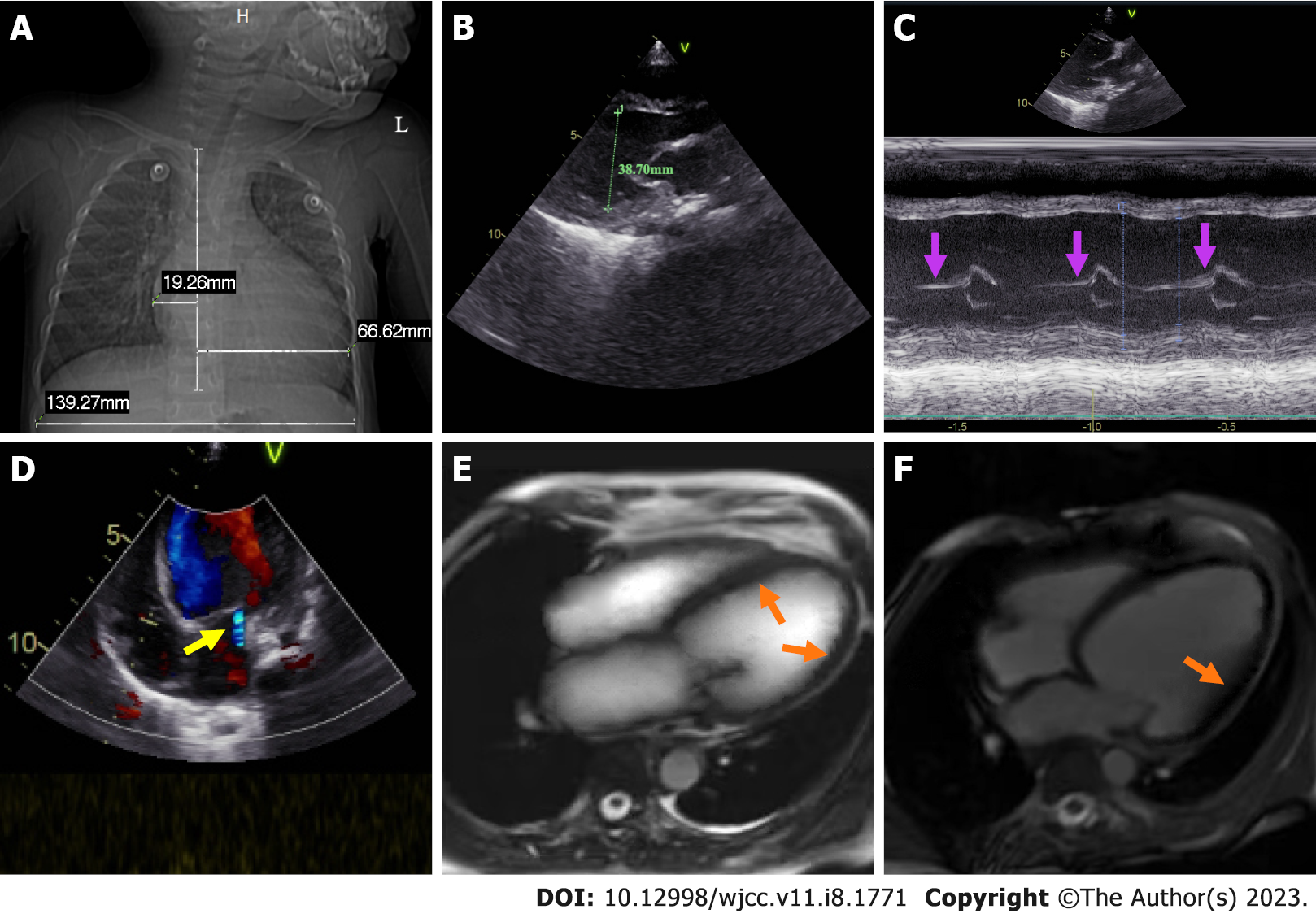
Figure 1 Chest radiograph, color doppler echocardiography, and cardiac magnetic resonance of the child.
A: Prominently enlarged heart with a high cardiothoracic ratio; B: Marked enlargement of the left ventricle (inner diameter 38.70 mm) with diffuse slight thickening of the endocardium; C: Thickening of the posterior wall of the left ventricle (purple arrow); D: A small amount of mitral regurgitation (yellow arrow); E: Bright-blood T1-weighted image shows slight endocardial thickening of the left ventricle and slight thickening of the myocardium (orange arrow); F: Dark-blood T2-weighted image shows diffuse ventricular wall hypokinesis without visible delayed enhancement abnormalities (orange arrow).
- Citation: Xie YY, Li QL, Li XL, Yang F. Pediatric acute heart failure caused by endocardial fibroelastosis mimicking dilated cardiomyopathy: A case report. World J Clin Cases 2023; 11(8): 1771-1781
- URL: https://www.wjgnet.com/2307-8960/full/v11/i8/1771.htm
- DOI: https://dx.doi.org/10.12998/wjcc.v11.i8.1771


