Copyright
©The Author(s) 2023.
World J Clin Cases. Mar 6, 2023; 11(7): 1656-1665
Published online Mar 6, 2023. doi: 10.12998/wjcc.v11.i7.1656
Published online Mar 6, 2023. doi: 10.12998/wjcc.v11.i7.1656
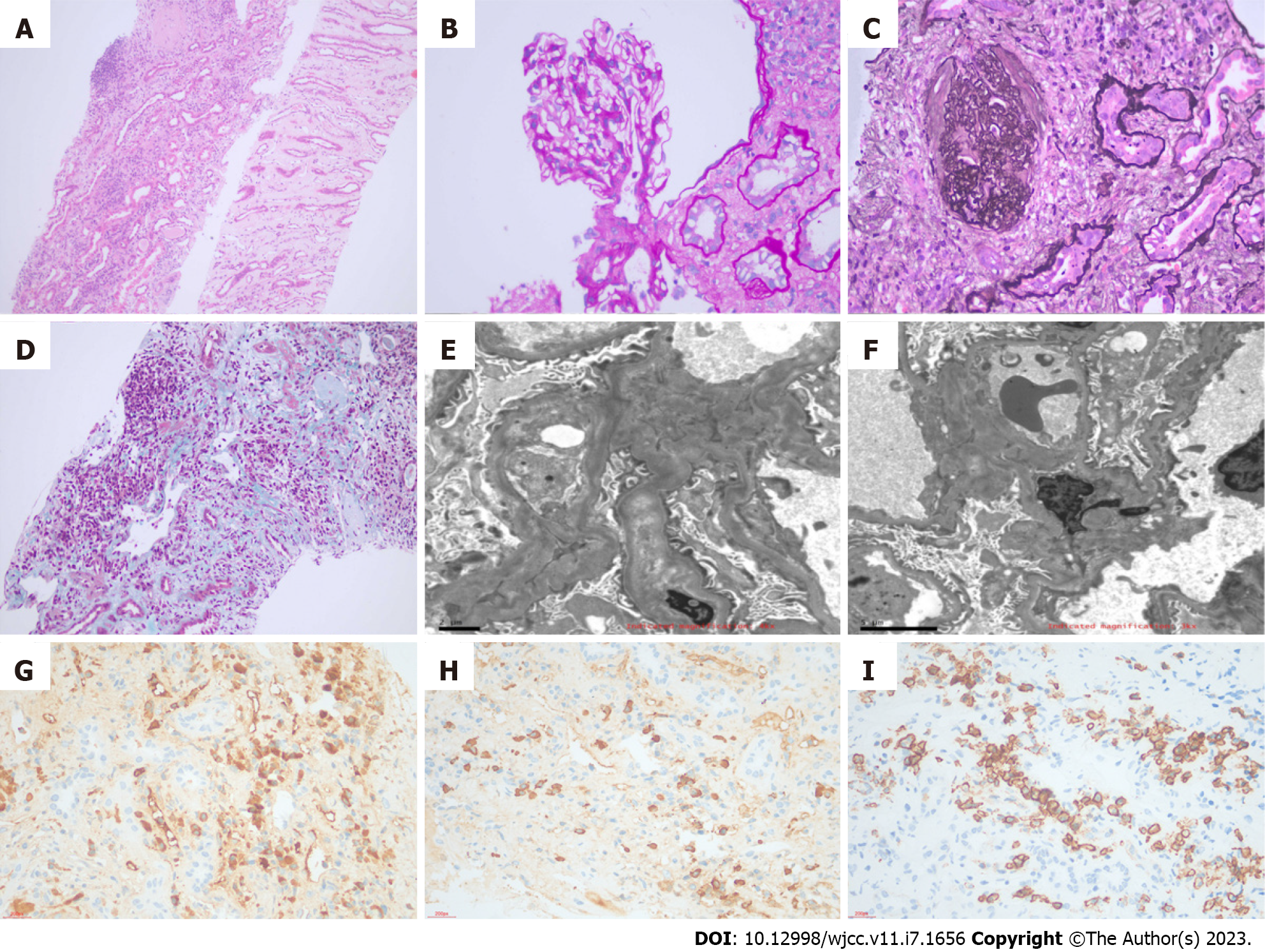
Figure 2 Histopathological findings on the kidney biopsy.
A: The renal interstitium was infiltrated by plasma cells and lymphocytes predominantly with fibrosis like car width [light microscopy: Hematoxylin and eosin (HE), × 100]; B: The injury of the glomerular lesion was mild [light microscopy: Periodic acid-Schiff staining, × 400]; C: Glomerular sclerosis and striate fibrosis in the renal interstitium [light microscopy: Masson and periodic acid-sliver methenamine, × 400]; D: Masson 200 staining of the lesion that was the same as that in Figure A; E and F: Most fusion of glomerular podocytes was observed by electron microscopy, while no electron dense granules were observed in the subepithelium, mesangial area, or subendothelium; G and H: IgG4 plasma cells > 10 cells/high powered field and IgG4 plasma cells/IgG plasma cells > 40% were observed by immunohistochemistry; I: There were CD38-positive plasma cells and CD138-positive plasma cells.
- Citation: He PH, Liu LC, Zhou XF, Xu JJ, Hong WH, Wang LC, Liu SJ, Zeng JH. IgG4-related kidney disease complicated with retroperitoneal fibrosis: A case report. World J Clin Cases 2023; 11(7): 1656-1665
- URL: https://www.wjgnet.com/2307-8960/full/v11/i7/1656.htm
- DOI: https://dx.doi.org/10.12998/wjcc.v11.i7.1656


